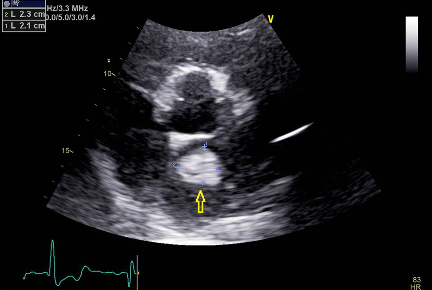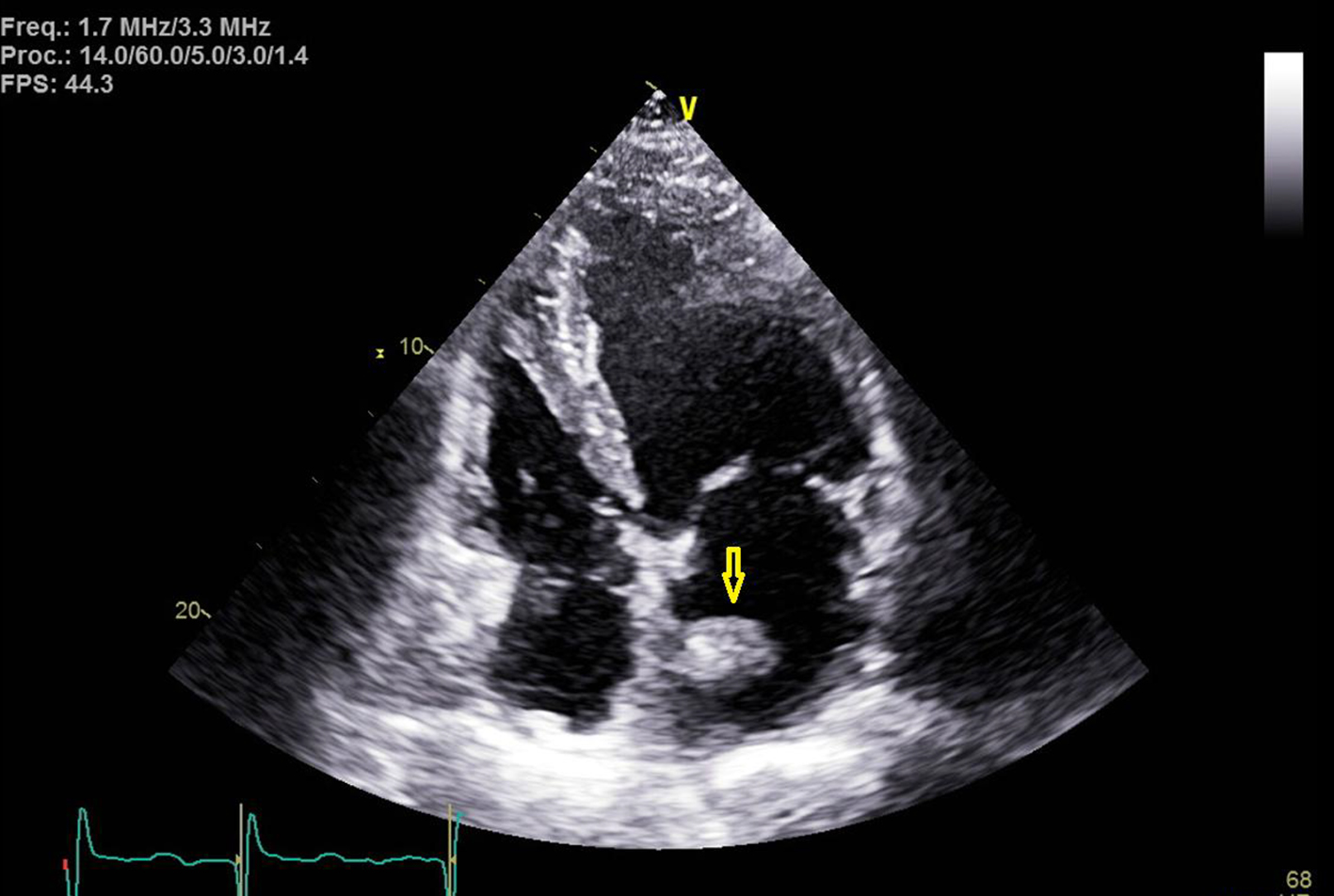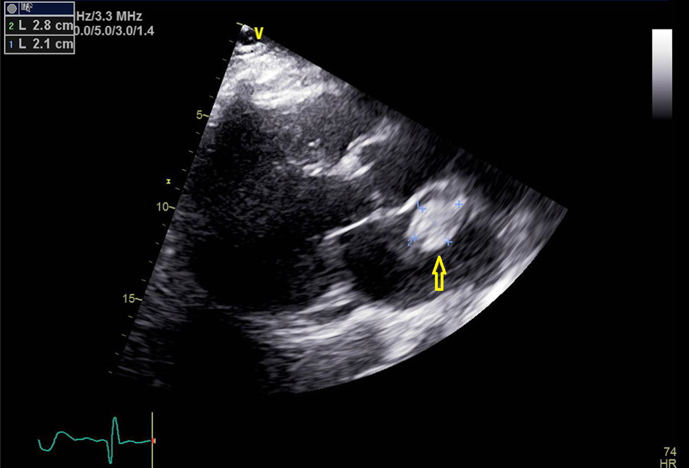
Figure 1. Transthoracic echocardiogram image with parasternal long axis view. Yellow arrow points towards the left atrial mass.
| Journal of Medical Cases, ISSN 1923-4155 print, 1923-4163 online, Open Access |
| Article copyright, the authors; Journal compilation copyright, J Med Cases and Elmer Press Inc |
| Journal website https://www.journalmc.org |
Case Report
Volume 12, Number 6, June 2021, pages 243-247
Left Atrial Thrombus Mimicking Myxoma Secondary to Rebound Hypercoagulable State
Figures