Journal of Medical Cases, ISSN 1923-4155 print, 1923-4163 online, Open Access
Article copyright, the authors; Journal compilation copyright, J Med Cases and Elmer Press Inc
Journal website https://www.journalmc.org

Case Report

Volume 12, Number 3, March 2021, pages 107-111


Immunoglobulin G4 Related-Disease: A Rare Presentation With Secondary Hypereosinophilic Syndrome and Eosinophilic Ascites

Figures

Figure 1.
Figure 1. Evolution of the peripheral blood absolute eosinophil count (normal range (light-gray): 40 - 400 cells/µL) during the disease course. Day 0 refers to admission day. The patient started corticosteroids on day 2 and was discharged on day 10. Thereafter, the patient was observed as an outpatient on days 13, 36, and 94, remaining stable despite progressive corticosteroid tapering.
Figure 2.
Figure 2. Upper digestive tract of the young female patient 2 days after starting the steroid therapy. In the superficial esophagus biopsy fragments obtained in the upper digestive tract endoscopy there was a light inflammatory infiltrate composed mainly by eosinophils, recognizable by their cytoplasmic granules that appear brick-red after eosin staining (yellow circles) with a maximum of five eosinophils per high-power field. (hematoxylin and eosin stain (H&E), × 200 magnification).
Figure 3.
Figure 3. Lower digestive tract of the young female patient 2 days after starting methylprednisolone. In the ileal biopsy fragments obtained in the lower digestive tract endoscopy the global architecture and mucosecretory activity were globally preserved, with the chorion containing a mild polymorphic inflammatory infiltrate rich in eosinophils, recognizable by their cytoplasmic granules that appear brick-red after eosin staining (yellow circles) (hematoxylin and eosin stain (H&E), × 200 magnification).

Table

Table 1. Immunology and Serology Laboratory Results of the Young Female Patient
 
PatientNormal range
Anti-dsDNA: anti-double stranded DNA; HAV: hepatitis A virus; HB: hepatitis B; HCV: hepatitis C virus; HIV: human immunodeficiency virus; HTLV: human T-cell lymphotropic virus.
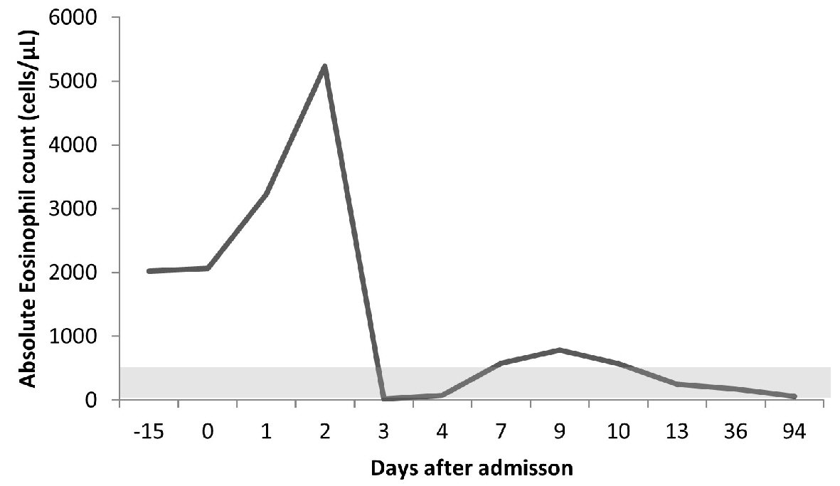
Immunoglobulins (Igs)
  IgA, mg/dL150.0114.0 - 457.0
  IgG, mg/dL1,061.0793.0 - 1,590.0
    IgG1, mg/dL778.0240 - 1,118
    IgG2, mg/dL267.0124 - 549
    IgG3, mg/dL44.221 - 134
    IgG4, mg/dL183.07 - 89
  IgM, mg/dL30.929.0 - 226.0
  IgE, KU/L13< 100
  Ig light-chains kappa, mg/dL247
  Ig light-chains lambda, mg/dL144
  Ratio kappa/lambda1.721.35 - 2.70
Complement (C)
  C3, mg/dL119.581.0 - 167.0
  C4, mg/dL12.011.0 - 42.0
Allergy
  PhadiatopNegative
  Tryptase, µg/L2.41< 11.4
Auto-antibodies (Ab)
  Anti-nuclear antibodies (ANAs)Negative
  Anti-dsDNA, UI/mL0.6< 15.0
  Anti-Sm, U/mL0.2< 10.0
  Anti-SS-A, U/mL0.2< 10
  Anti-SS-B, U/mL0.1< 10
  IgA anti-transglutaminase, U/mL0.3< 10
  Antineutrophil cytoplasmic antibodies (ANCAs)Negative< 1/20
  Anti-proteinase 3 (PR3), UQ< 2.3< 20
  Anti-myeloperoxidase (MPO), UQ4.1< 20
  IgA anti-Saccharomyces cerevisiae antibody (ASCA), U/mL1.7< 10
  IgG anti-Saccharomyces cerevisiae antibody (ASCA), U/mL0.4<10
Serology
  Anti-Treponema pallidum AbNegative
  Anti-HAV AbNegative
  Antigen HBsNegative
  Anti-HBs Ab, UI/LPositive (94.1)
  Anti-HBc AbNegative
  Anti-HCV AbNegative
  Anti-HIV AbNegative
  Anti-HTLV INegative
  Anti-HTLV INegative
  Fasciola hepatica< 1/160Negative: < 1/160
  Toxocara canisNegative
  SchistosomaNegative
  Echinococcus IgG index0.7Negative: < 9
  Strongyloides stercoralis AbNegative
  Ascaris, kUA/L0.01< 0.35