Journal of Medical Cases, ISSN 1923-4155 print, 1923-4163 online, Open Access
Article copyright, the authors; Journal compilation copyright, J Med Cases and Elmer Press Inc
Journal website https://www.journalmc.org

Case Report

Volume 12, Number 2, February 2021, pages 57-60


Transvaginal Resection of a Rectal Gastrointestinal Stromal Tumor: A Case Report

Figures

Figure 1.
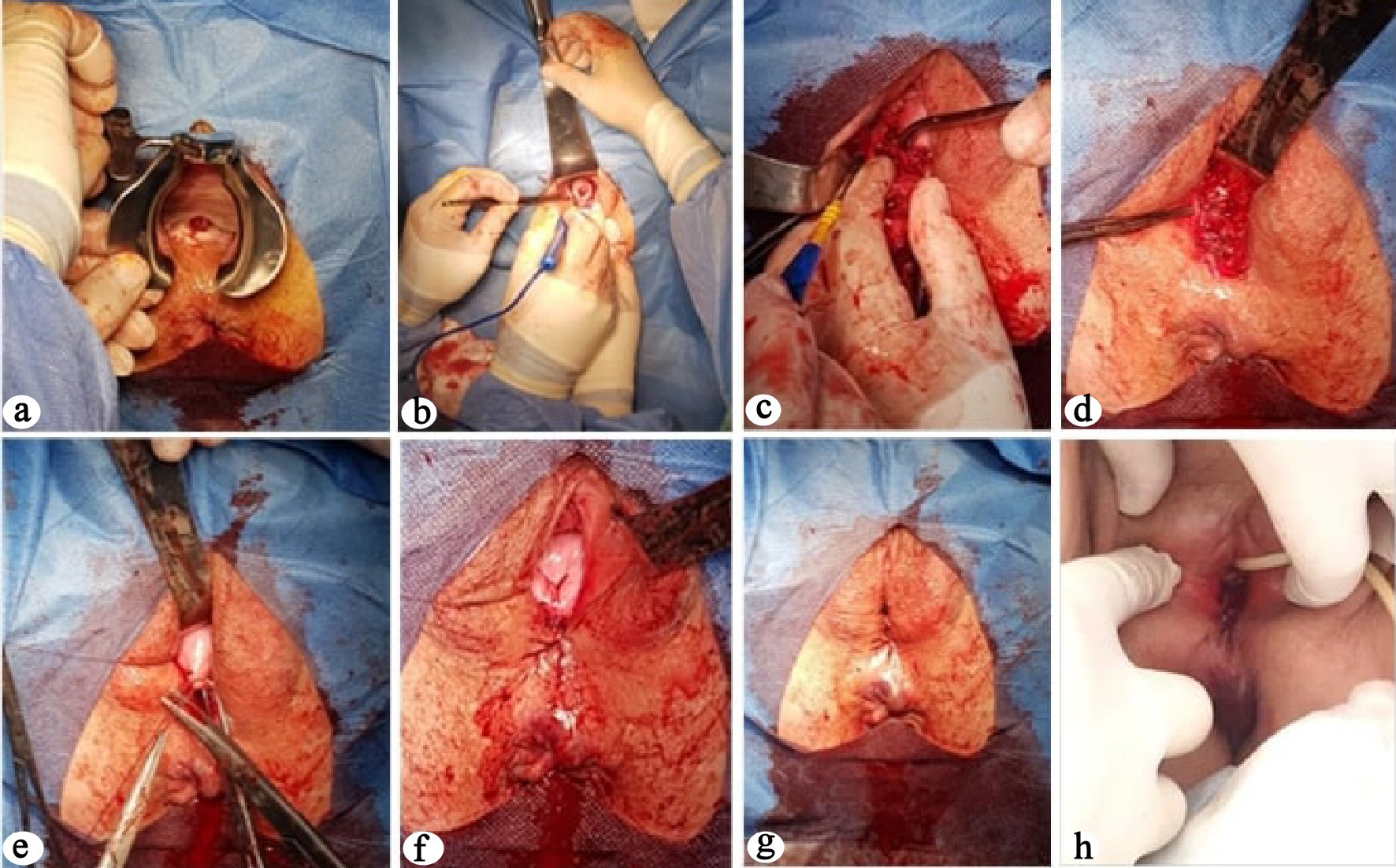
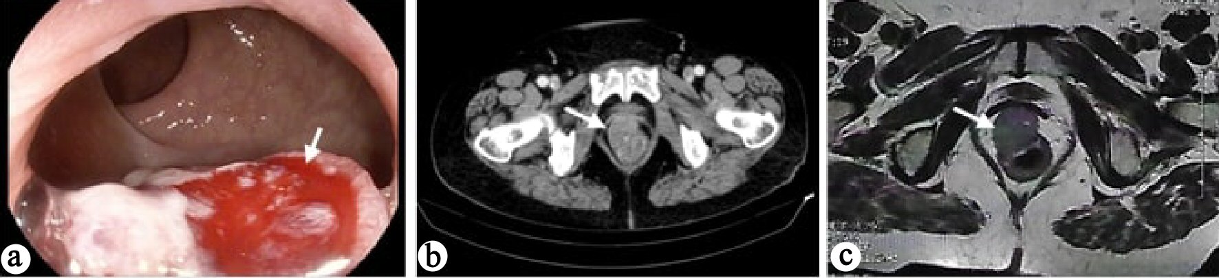
Figure 1. Complementary diagnostic tests. (a) Colonoscopy: ulcerated, hard and friable lesion (arrow), located on the anterior surface of the distal anal-rectal rectum. (b) CT scan: tumor formation (arrow) in the rectum, with no densification of the fat of the ischiorectal space or lymphadenopathy. (c) Pelvic MRI scan: mass (arrow) in the rectovaginal space of 5.1 × 3.1 cm with origin in the anterior wall of the rectum about 2.9 cm from the anal canal with presumably non-epithelial origin and without apparent local invasion.
Figure 2.
Figure 2. Surgical intervention: transvaginal resection. (a) Protrusion of the lesion through the posterior wall of the vagina. (b-d) Transvaginal excision of the lesion. (e-g) Defect closure by layers. (h) The first postoperative day.