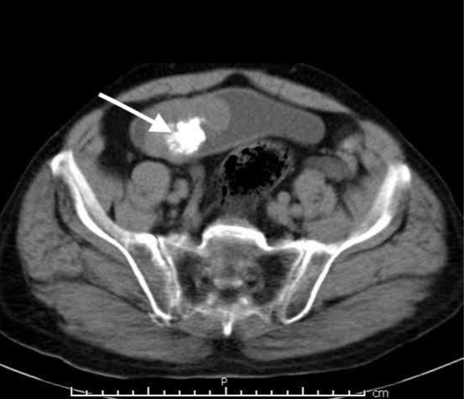
Figure 1. Computed tomography findings of the urinary bladder mass lesion (arrow denotes the tumor).
| Journal of Medical Cases, ISSN 1923-4155 print, 1923-4163 online, Open Access |
| Article copyright, the authors; Journal compilation copyright, J Med Cases and Elmer Press Inc |
| Journal website https://www.journalmc.org |
Case Report
Volume 12, Number 7, July 2021, pages 280-283
Primary Osteosarcoma of the Urinary Bladder Metastatic to Lung
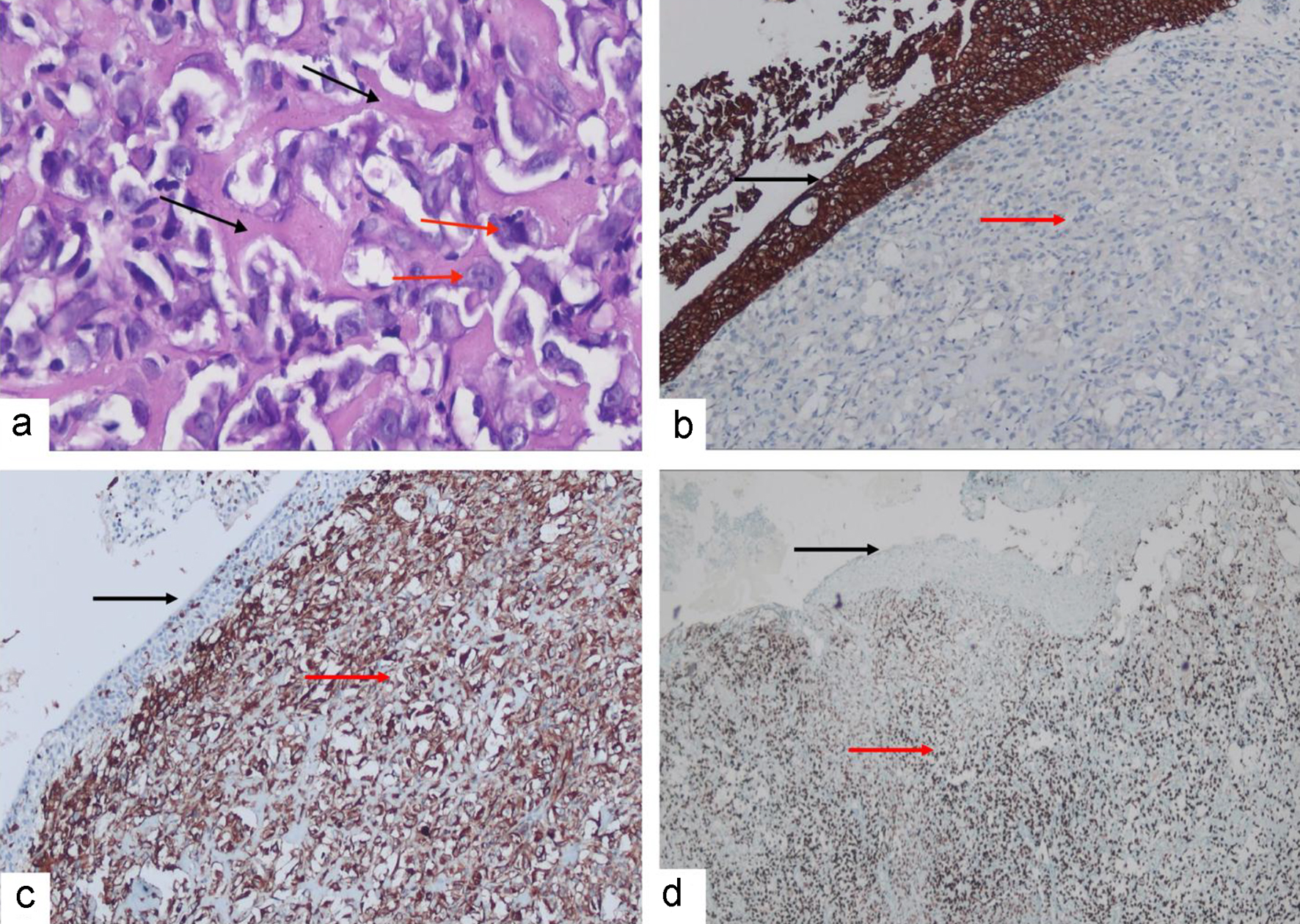
Figures