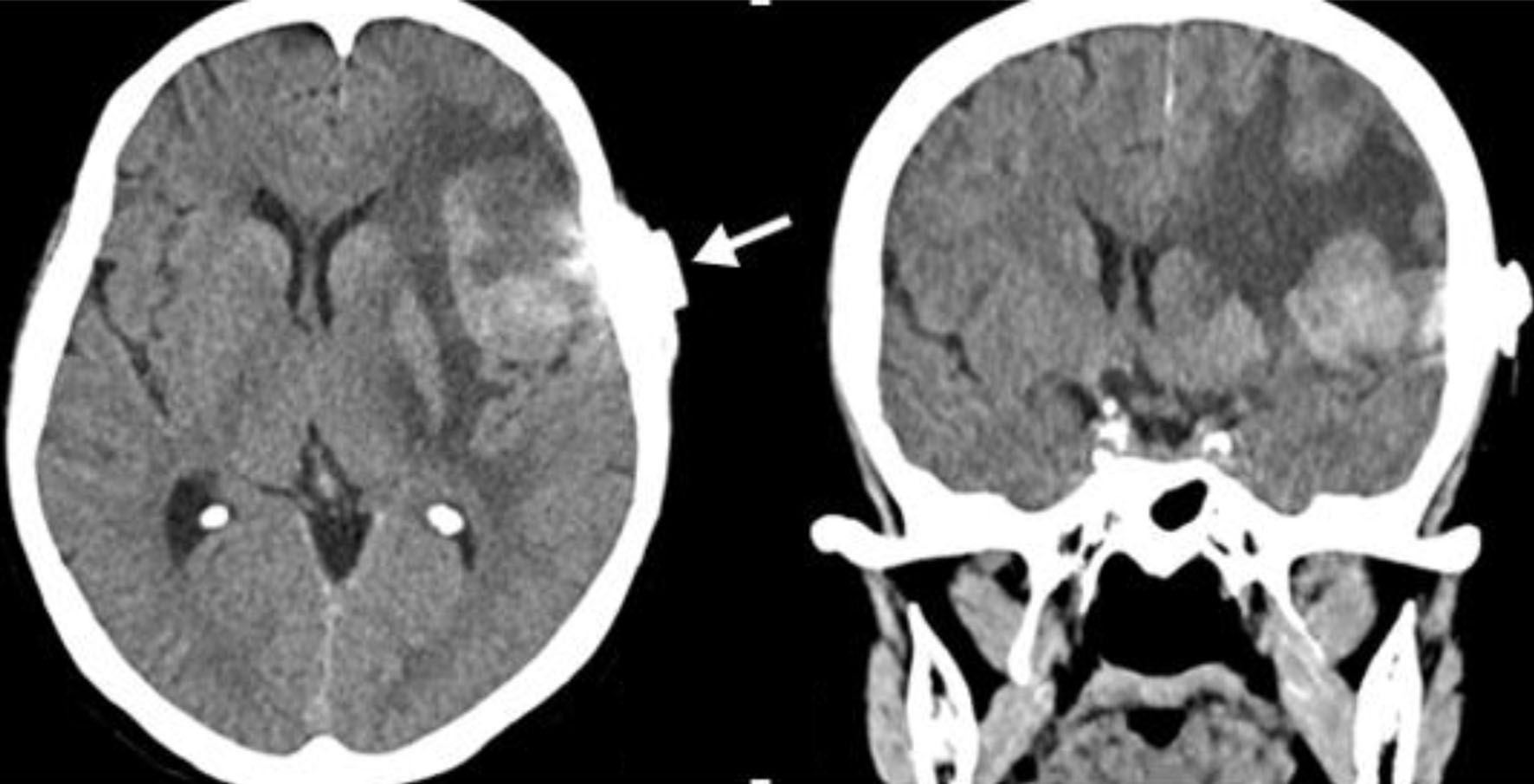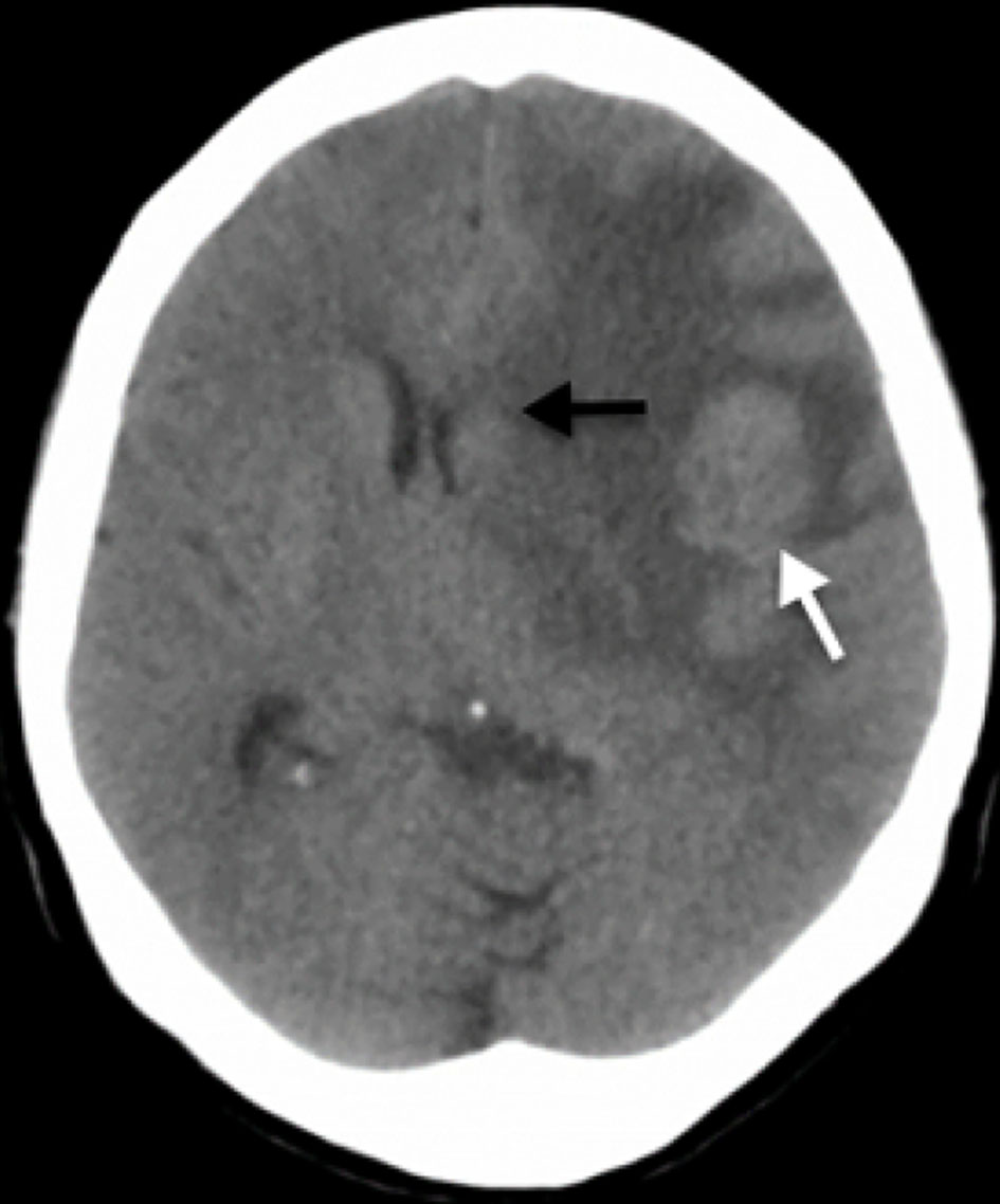
Figure 1. Simple skull CT scan. A rounded lesion is seen in the middle portion of the subcortical cortical area in the left frontal lobe (white arrow) with vasogenic edema that together, generate a mass effect with deviation of the midline to the right, compression of lateral ventricles and subfalcine herniation (black arrow). CT: computed tomography.
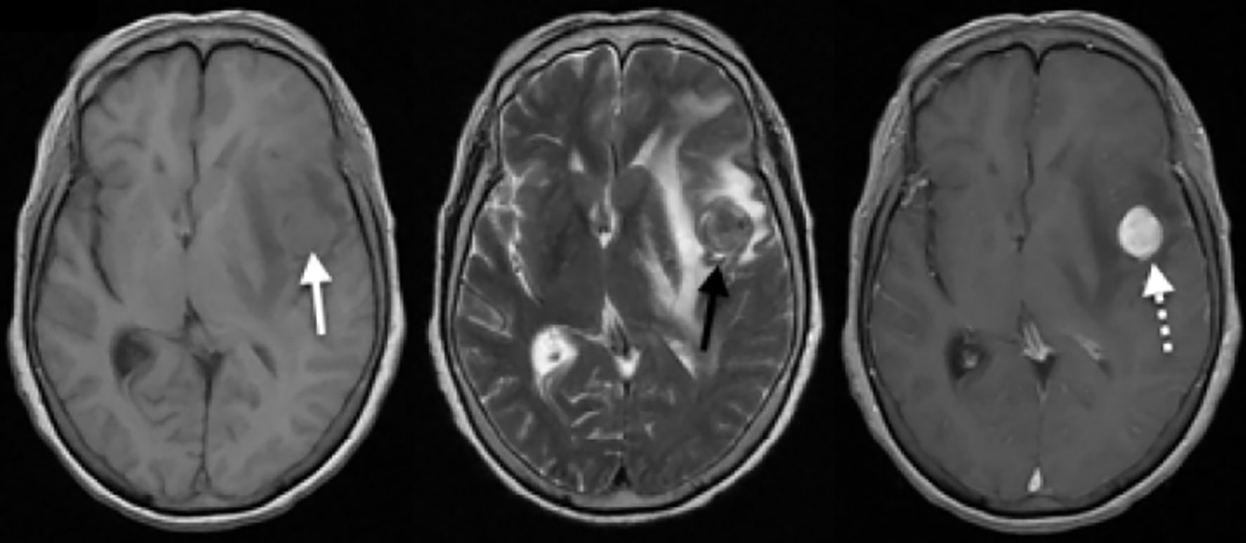
Figure 2. Simple and contrasted brain MRI. A homogeneous isointense rounded lesion in T1 is seen in the medical subcortical cortical region of the left frontal lobe (white arrow), with associated vasogenic edema and compressive effect on the lateral ventricles, with deviation of the midline to the right. For the T2 sequence, heterogeneous densities resembled its interior with a hypointense component and the extent of digitiform edema is seen in better detail (black arrow). After administration of the contrast medium, a homogeneous uptake of the entire lesion is observed (white dashed arrow), highlighting its rounded and circumscribed shape. MRI: magnetic resonance imaging.