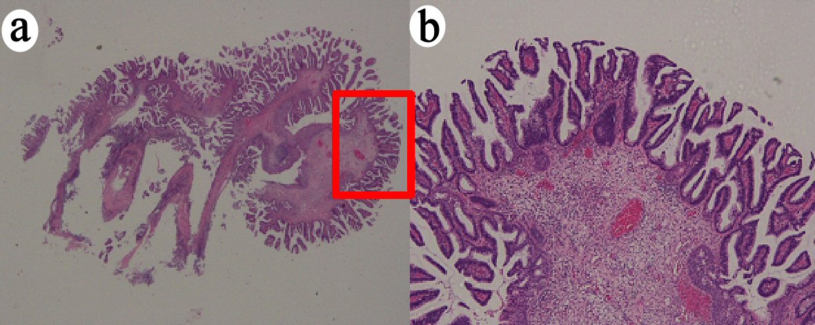
Figure 1. VGA obtained by polypectomy. (a) Whole mounted view of cervical exophytic tumor (hematoxylin and eosin (H&E) stain, × 1). (b) The papillae are short, covered by atypical endocervical epithelia, in a villoglandular lesion (H&E stain, × 4). VGA: villoglandular papillary adenocarcinoma.
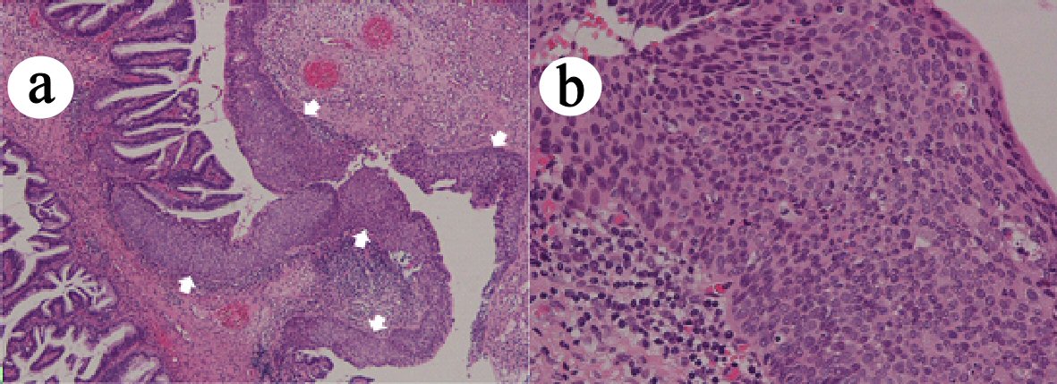
Figure 2. CIN3, coexisted with adenocarcinoma. (a) CIN3 lesion (indicated by white arrows) existed with adenocarcinoma (hematoxylin and eosin (H&E) stain, × 4). (b) CIN3 lesion shows severe atypia reaching the upper third of the squamous epithelium (H&E stain, × 20). CIN: cervical intraepithelial neoplasia.
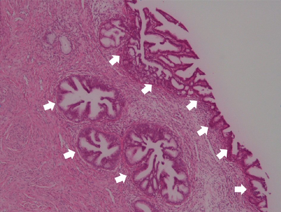
Figure 3. Residual AIS lesion. Residual AIS lesion (indicated by white arrows) that appeared to correspond to the basal part of VGA was observed after cone biopsy (hematoxylin and eosin (H&E) stain, × 4). AIS: adenocarcinoma in situ; VGA: villoglandular papillary adenocarcinoma.