Journal of Medical Cases, ISSN 1923-4155 print, 1923-4163 online, Open Access
Article copyright, the authors; Journal compilation copyright, J Med Cases and Elmer Press Inc
Journal website https://www.journalmc.org

Case Report

Volume 11, Number 12, December 2020, pages 400-402


COVID-19 Pneumonia or Hypereosinophilic Syndrome?

Figures

Figure 1.
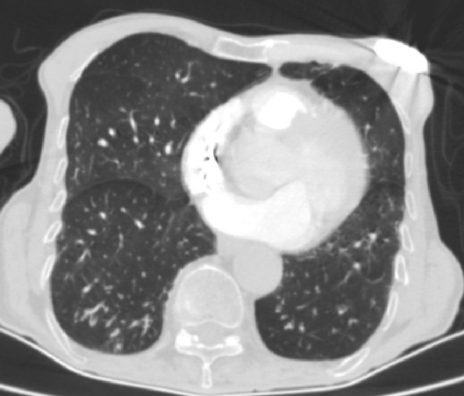
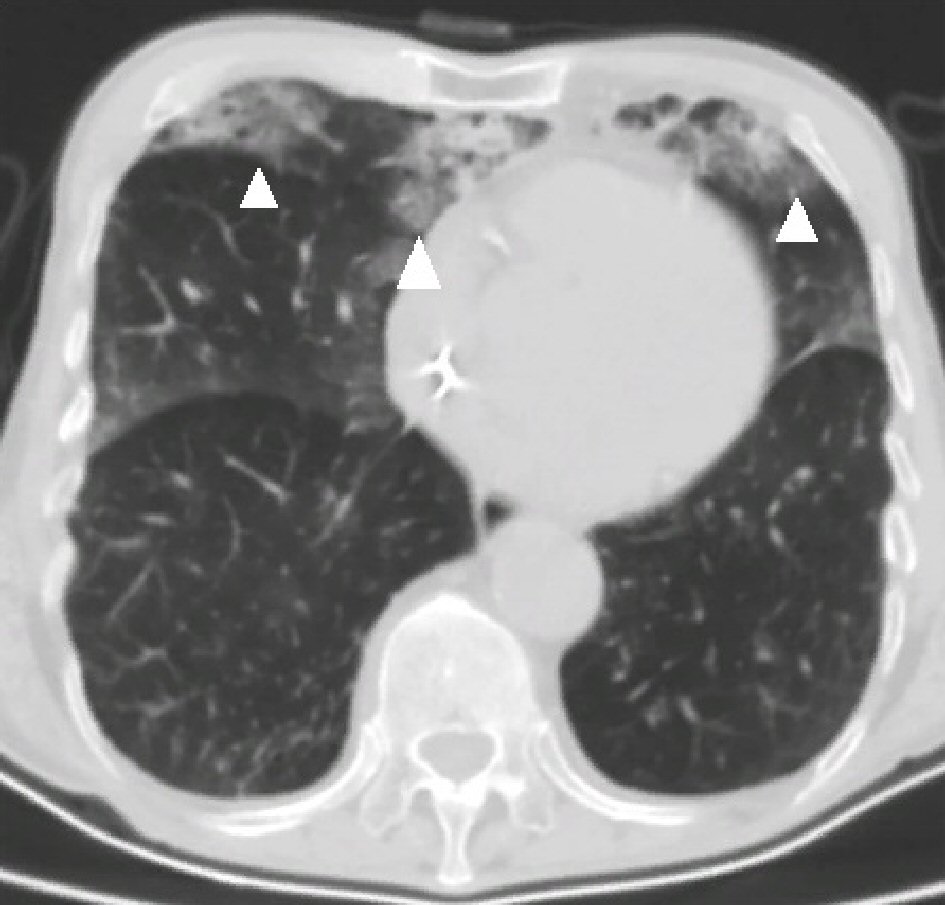
Figure 1. Pulmonary involvement of HES, computed tomography at initial diagnosis: patchy images of HES involvement (arrows). There was no pathological lymphadenomegaly in the mediastinum. In the evaluation of the lung parenchyma structures, ground glass patchy consolidation areas were observed at the level of the middle and lower lobe segments. Ground glass densities were determined at the level of the upper lobe segments, compatible with proliferative phase COVID-19 pneumonia. HES: Hypereosinophilic syndrome; COVID-19: coronavirus disease 2019.
Figure 2.
Figure 2. Three Weeks after beginning of treatment: complete regression. It appears that all visible involvement ground glass patchy areas have disappeared 3 weeks after beginning of treatment.