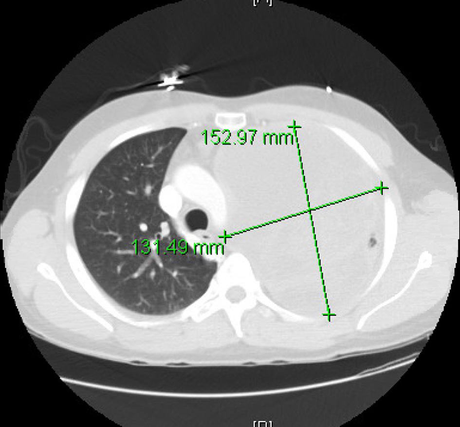
Figure 1. Computed tomography (CT) thorax showing left lung mass with a left to right mediastinal shift.
| Journal of Medical Cases, ISSN 1923-4155 print, 1923-4163 online, Open Access |
| Article copyright, the authors; Journal compilation copyright, J Med Cases and Elmer Press Inc |
| Journal website https://www.journalmc.org |
Case Report
Volume 11, Number 12, December 2020, pages 388-393
Local Lung Mass Masquerading a Very Aggressive Extraskeletal Ewing Sarcoma Presenting as Bilateral Paraparesis in a Young Adult
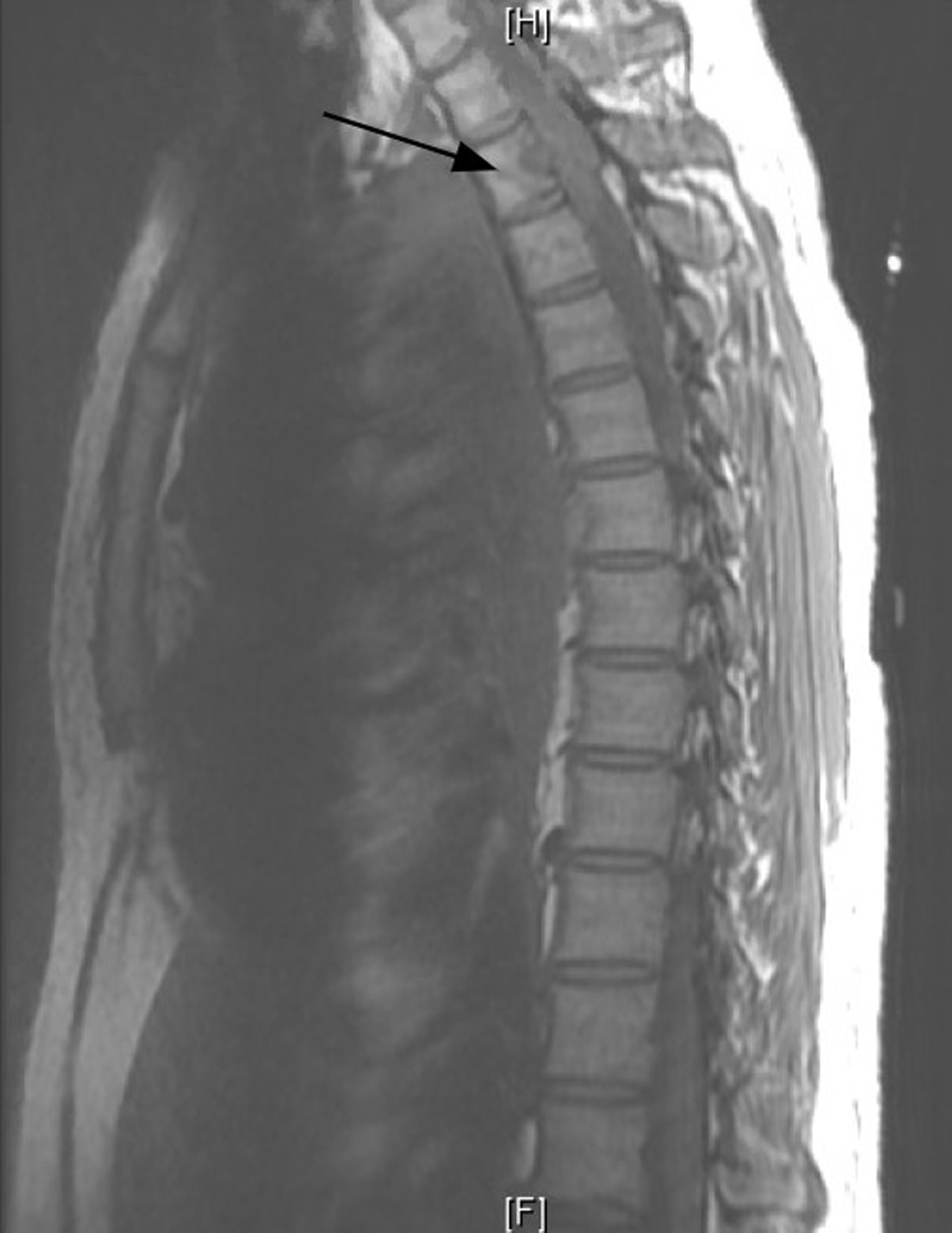
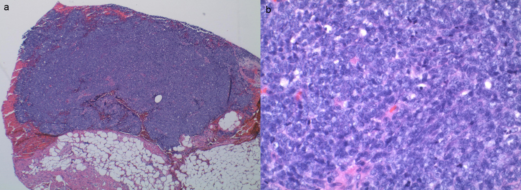
Figures
Table
| Laboratory | Result | Normal value |
|---|---|---|
| White blood cells | 21.07 × 10³/µL | 4 - 10 × 103/µL |
| Hemoglobin | 13.2 g/dL | 11.2 - 15.7 g/dL |
| Platelets | 418 × 103/µL | 163 - 369 × 103/µL |
| Sodium | 143 mEq/L | 136 - 144 mEq/L |
| Potassium | 4.0 mEq/L | 3.5 - 5.1 mEq/L |
| Chloride | 103 mEq/L | 98 - 110 mEq/L |
| Bicarbonate | 26 mEq/L | 20 - 30 mEq/L |
| Blood urea nitrogen | 12 mg/dL | 7 - 23 mg/dL |
| Creatinine | 0.84 mg/dL | 0.57 - 1.11 mg/dL |
| Aspartate transaminase | 19 U/L | 5 - 42 U/L |
| Alanine transaminase | 35 U/L | 5 - 49 U/L |
| Alkaline phosphatase | 73 U/L | 35 - 141 U/L |
| Total bilirubin | 0.3 mg/dL | 0.1 - 1.2 mg/dL |