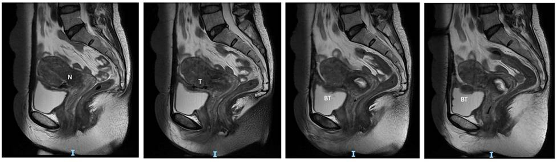
Figure 1. MRI images showing the sagittal view of the Cesarean scar niche (N), tract (T) and bladder tumor (BT). MRI: magnetic resonance imaging.
| Journal of Medical Cases, ISSN 1923-4155 print, 1923-4163 online, Open Access |
| Article copyright, the authors; Journal compilation copyright, J Med Cases and Elmer Press Inc |
| Journal website https://www.journalmc.org |
Case Report
Volume 11, Number 11, November 2020, pages 370-373
Isolated Bladder Endometriosis in a Patient With Previous Cesarean Sections
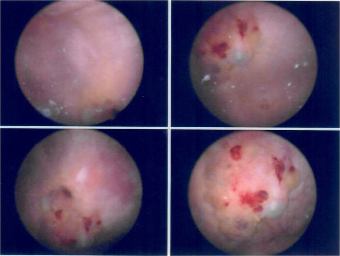
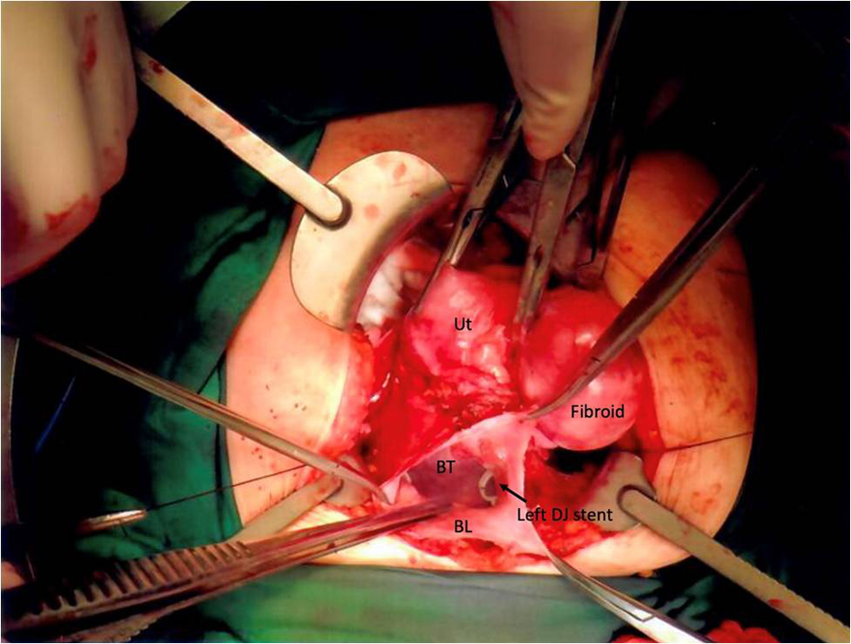
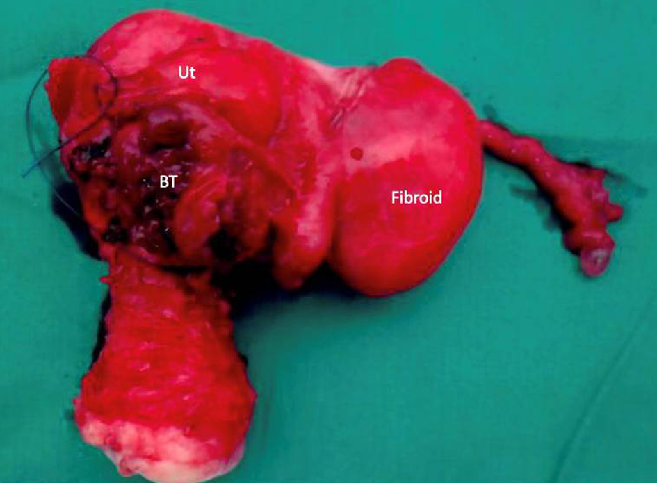
Figures