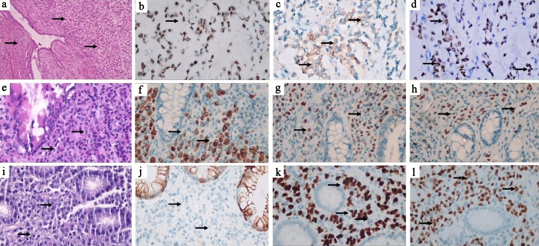
Figure 1. Case 1: (a) Hematoxylin and eosin (H&E) stained section showing infiltration of the cervical wall by non-cohesive tumor cells (black arrows), × 100. (b) Tumor cells show nuclear reaction for GATA3 (black arrow) × 400. (c) Tumor cells show cytoplasmic reaction for mammaglobin (black arrows) × 400 and (d) nuclear reaction for ER (black arrows) × 400. Case 2: (e) H&E stained sections of colonic mucosa diffusely infiltrated by tumor cells (× 100). (f) Tumor cells show cytoplasmic reaction for CK7 (black arrows), × 400. (g) Nuclear reaction for ER (black arrows), × 400 and (h) GATA-3 (black arrow), × 400. Case 3: (i) H&E stained section showing infiltration of the cervical wall by non-cohesive tumor cells (black arrows), × 400. (j) Tumor cells are negative for CK20 (black arrows), which stains the surface mucosa, × 400. (k) Tumor cells show positive nuclear reaction for ER (black arrows), × 400 and (l) GATA-3 (black arrows), × 400. CK: cytokeratin; ER: estrogen receptor.