Journal of Medical Cases, ISSN 1923-4155 print, 1923-4163 online, Open Access
Article copyright, the authors; Journal compilation copyright, J Med Cases and Elmer Press Inc
Journal website http://www.journalmc.org

Case Report

Volume 11, Number 8, August 2020, pages 249-252


De Novo Granulocytic Sarcoma of the Humerus in an 82-Year-Old Woman: A Case Report

Figures

Figure 1.
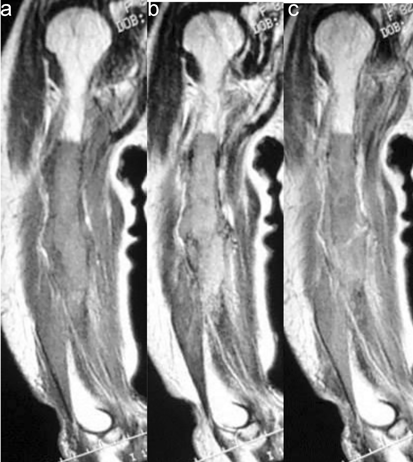
Figure 1. (a) Coronal T1-weighted MR image of the right humerus demonstrated both intra- and extramedullary lesion with low signal intensity. (b) Coronal T2-weighted MR image revealed a lesion with high signal intensity. (c) The lesion showed irregular and moderate enhancement after administration of gadolinium. MR: magnetic resonance.
Figure 2.
Figure 2. Positron emission tomography with 18F-fluorodeoxyglucose (FDG-PET) demonstrated the high maximum standardized uptake value (SUVmax) in the right humerus and bilateral thyroid glands.
Figure 3.
Figure 3. Photomicrograph of the immunostained humerus tumor showed CD68-positive tumor cells.