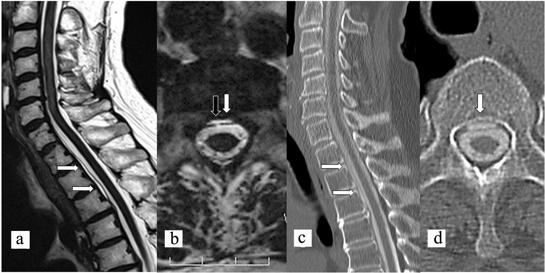
Figure 1. Preoperative sagittal (a) and axial (b) CISS MR images showing fluid collection (white arrows) in the spinal canal and a dural defect (black arrow). Preoperative sagittal (c) and axial (d) CT myelography showing longitudinal epidural CSF leakage (white arrows) ventral to the spinal cord. CISS: constructive interference in steady state; MR: magnetic resonance; CT: computed tomography; CSF: cerebrospinal fluid.
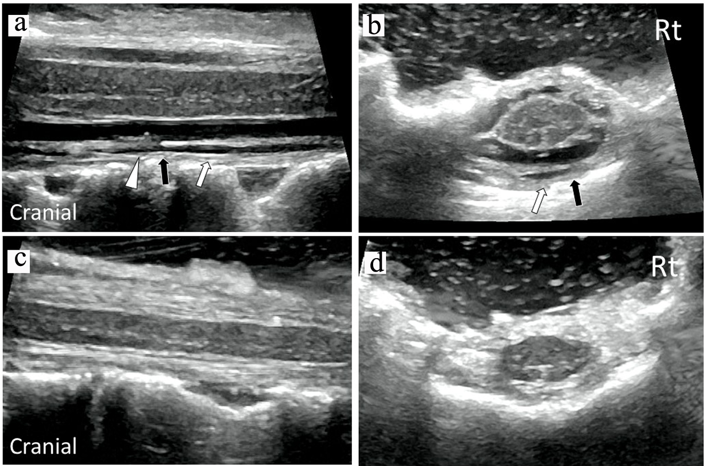
Figure 2. Intraoperative sagittal (a) and axial (b) US images confirmed verrucous vegetation (white triangle) and epidural fluid collection (white arrows) around the ventral dural defect (black arrow) before dural closure. Intraoperative sagittal (c) and axial (d) US images demonstrated removal of vegetation and resolution of fluid collection after dural closure. US: ultrasonography.
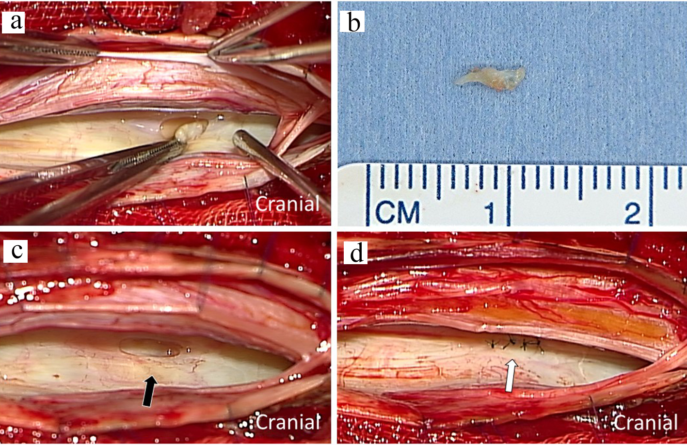
Figure 3. Intraoperative photograph showing dural defect with verrucous vegetation after incision of the posterior dura matter (a). The macroscopic image of resected specimen with scale (b). The ventral dural hole (black arrow) was detected during surgery (c). The dural defect was repaired by direct sutures (white arrows) (d).