Figures
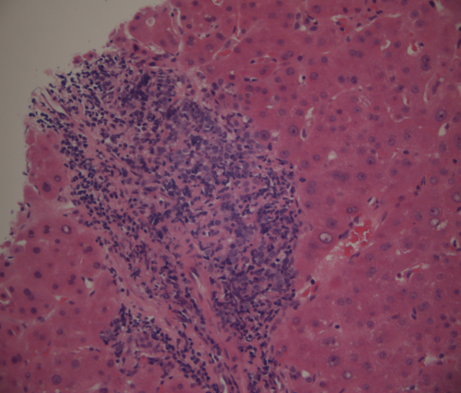
Figure 1. Low power view of portal tract lymphoma stained with haematoxylin and eosin.
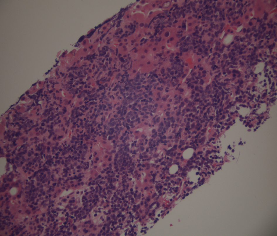
Figure 2. Low power view of liver parenchymal lymphoma stained with haematoxylin and eosin.
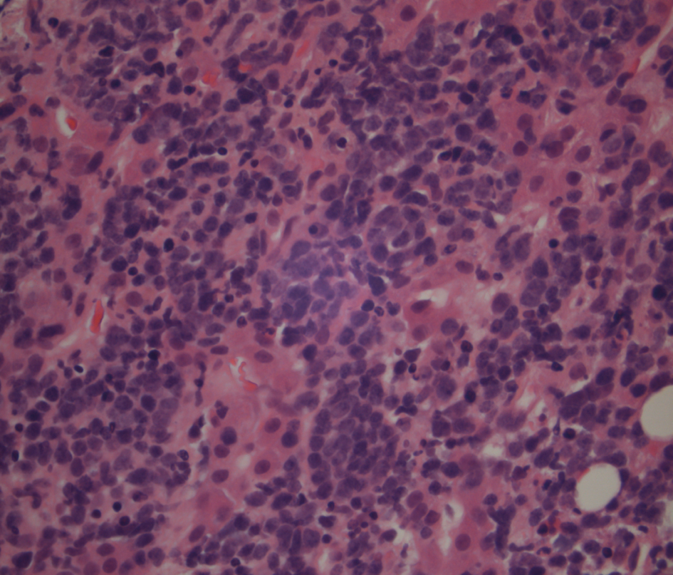
Figure 3. Liver biopsy showing a higher power of the lymphoma cells which have hyperchromatic nuclei and little cytoplasm.
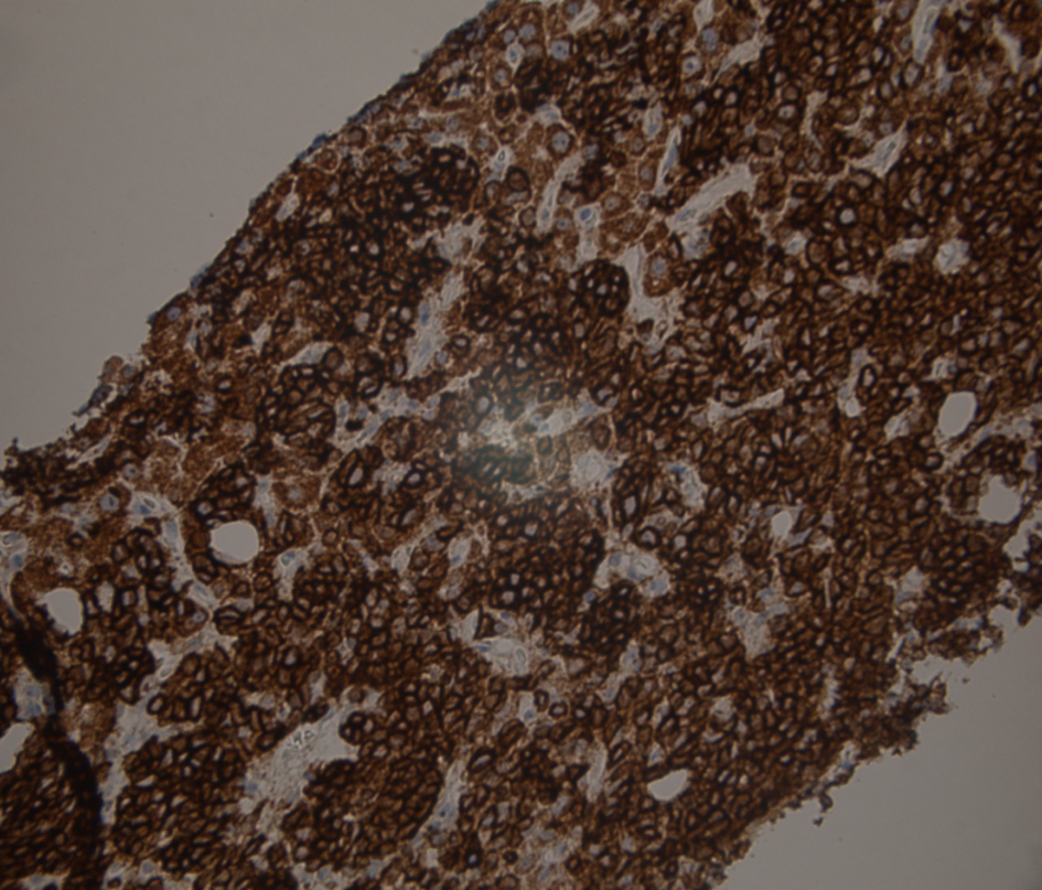
Figure 4. Liver biopsy immunostained for CD20, the usual B lymphocyte marker. Shows strong membrane and cytoplasmic positivity confirms that this is a B cell lymphoid proliferation.
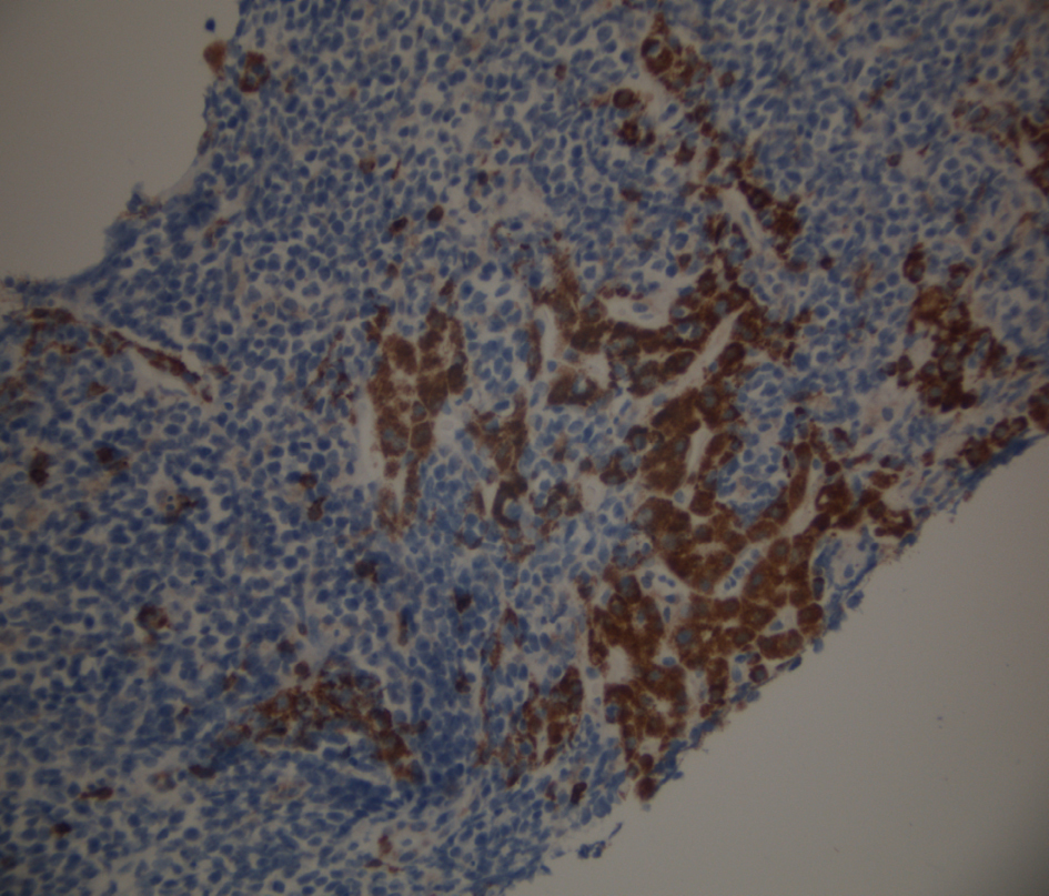
Figure 5. Liver biopsy stained for CD3, a T cell marker which is negative in the tumor cells. There is aberrant staining in hepatocytes due to incomplete blocking of endogenous peroxidases in the cells.
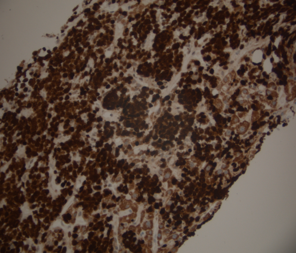
Figure 6. Liver biopsy stained for Mib-1 (Ki 67), a proliferation marker which is positive in the nuclei of cells in the mitotic cycle. In this case the proliferation index is nearly 100%, a finding associated with very aggressive lymphomas including Burkitts lymphoma and other diffuse large B cell lymphomas.