Figure 1. LAO cranial view showing right coronary artery dividing into PDA and PLV branches. LAO: left anterior oblique; PDA: posterior descending artery; PLV: posterior left ventricular.
| Journal of Medical Cases, ISSN 1923-4155 print, 1923-4163 online, Open Access |
| Article copyright, the authors; Journal compilation copyright, J Med Cases and Elmer Press Inc |
| Journal website http://www.journalmc.org |
Case Report
Volume 11, Number 4, April 2020, pages 97-99
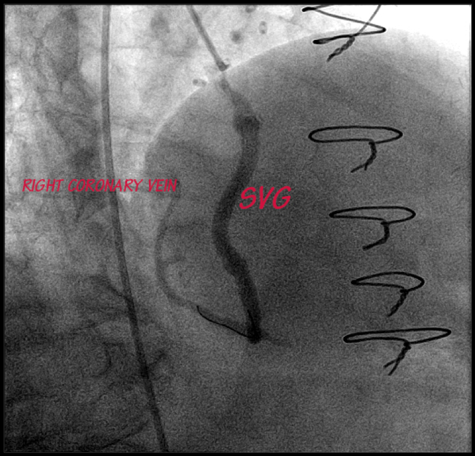
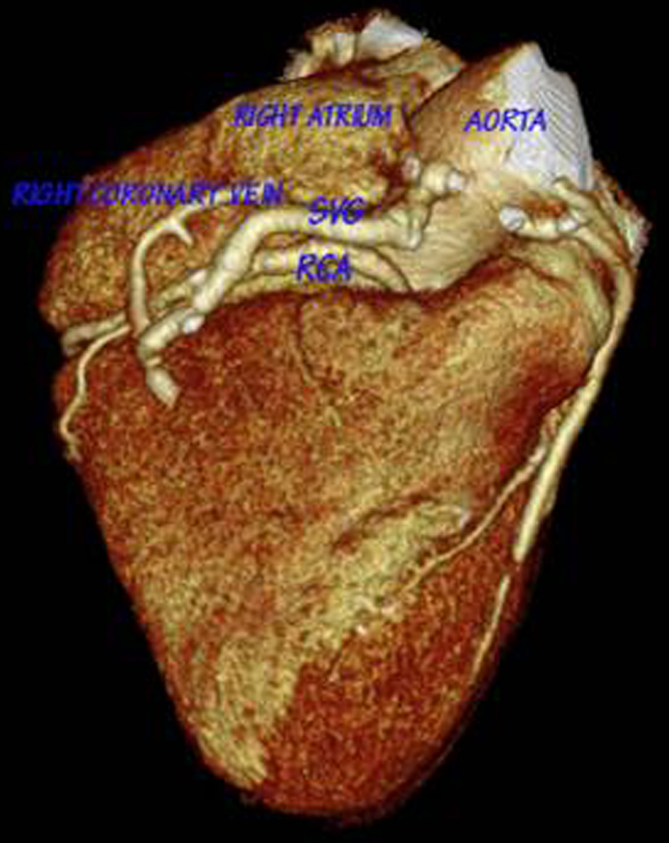
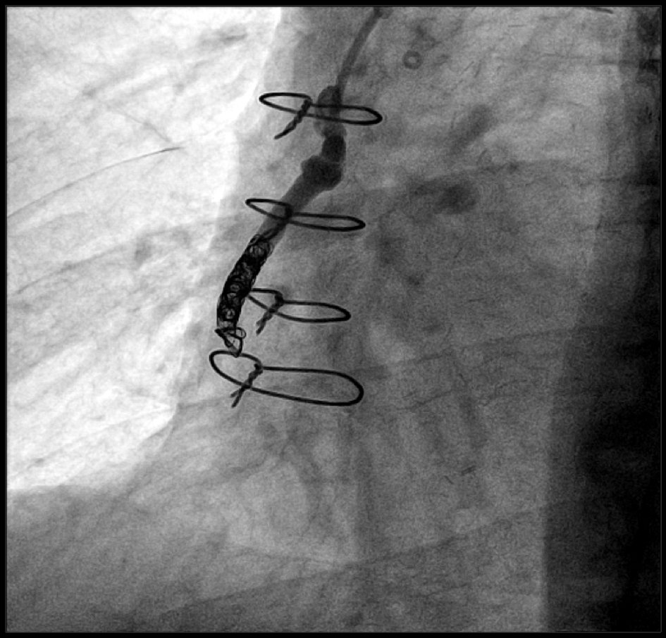
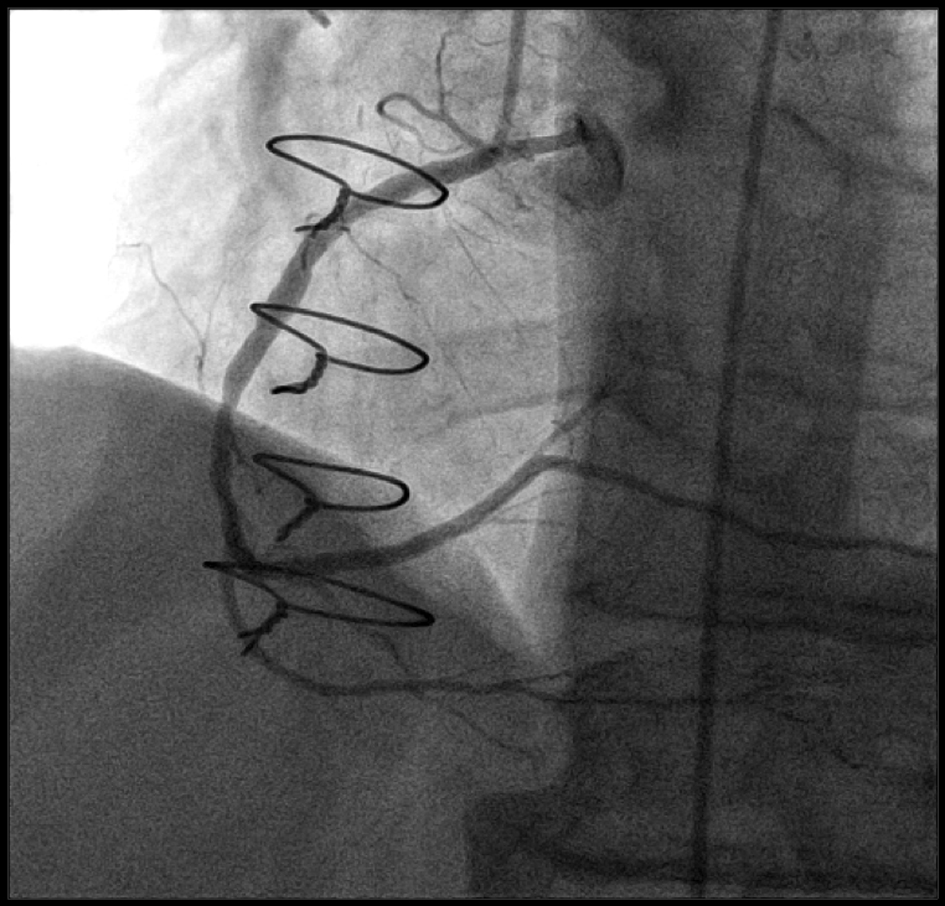
Transcatheter Coil Embolization of Iatrogenic Aorto-Right Atrial Fistula
Figures