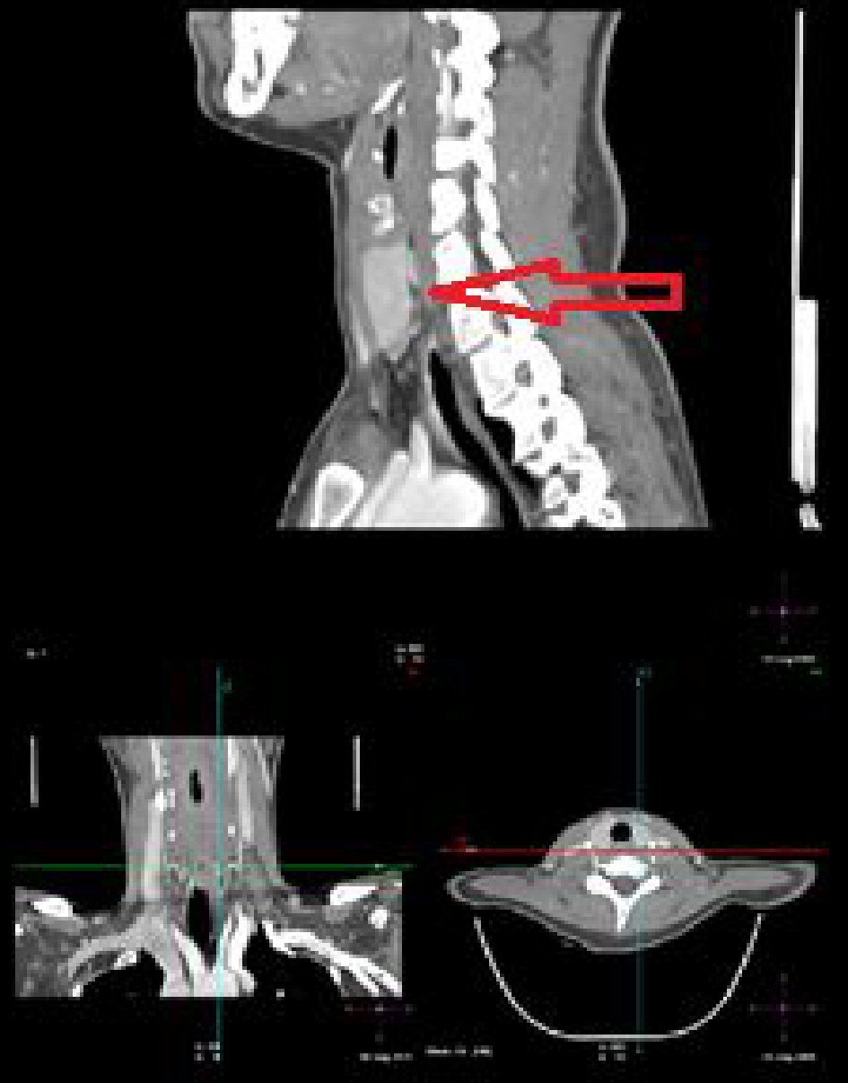
Figure 1. Computed tomography (CT) images of parathyroid adenoma (arrow).
| Journal of Medical Cases, ISSN 1923-4155 print, 1923-4163 online, Open Access |
| Article copyright, the authors; Journal compilation copyright, J Med Cases and Elmer Press Inc |
| Journal website http://www.journalmc.org |
Case Report
Volume 11, Number 4, April 2020, pages 83-85
Primary Hyperparathyroidism in Pregnancy: A Case Report
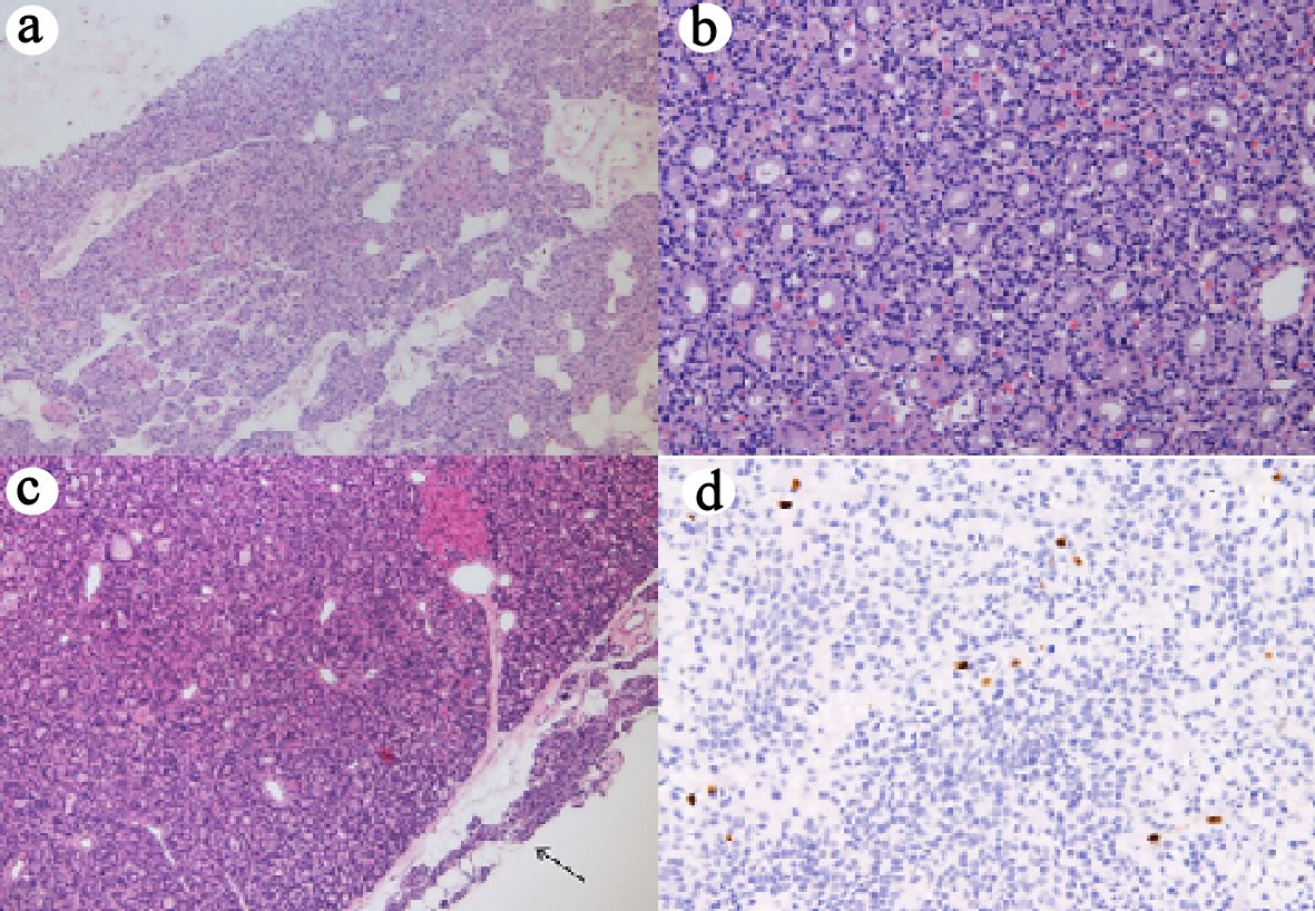
Figures