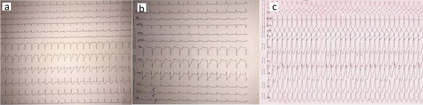
Figure 1. (a) Electrocardiogram showing normal sinus rhythm with first degree AV block, ST-segment elevation in II, III, and aVF; (b) Electrocardiogram showing ST-segment elevation in posterior leads V7 - V9; (c) Electrocardiogram showing recurrent monomorphic VT. AV: atrioventricular; VT: ventricular tachycardia.
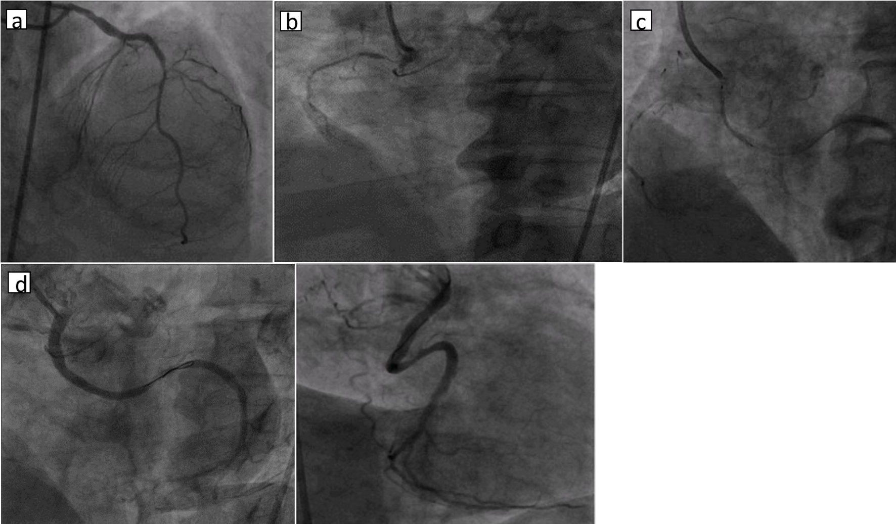
Figure 2. (a) Left coronary angiography showing normal LMCA and LAD; (b) right coronary angiography showing anamalous LCX from right sinus with total occlusion and non-dominant RCA; (c) right coronary angiography showing 3.5 × 24 mm DES deployed at 11 atm; (d) right coronary angiography showing dominant LCX with TIMI III flow (LAO and RAO views). LMCA: left main coronary artery; LAD: left anterior descending artery; LCX: left circumflex artery; DES: drug-eluting stent; LAO: left anterior oblique; RAO: right anterior oblique.