Journal of Medical Cases, ISSN 1923-4155 print, 1923-4163 online, Open Access
Article copyright, the authors; Journal compilation copyright, J Med Cases and Elmer Press Inc
Journal website http://www.journalmc.org

Case Report

Volume 10, Number 12, December 2019, pages 354-358


Salivary Duct Carcinoma of the Submandibular Gland: A Case Report and Review of the Literature

Figures

Figure 1.
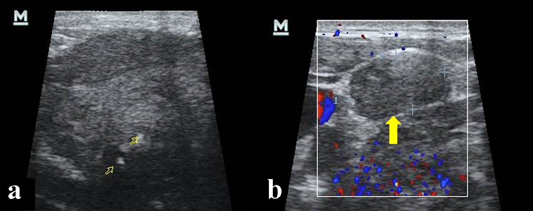
Figure 1. Cervical ultrasonography. (a) Hypoecogenic, heterogeneous solid mass and calcium deposit within (open yellow arrows). (b) Lymphadenopathy in V ganglionic level (yellow arrow).
Figure 2.
Figure 2. Computed tomography (CT) showing a primary left submandibulary mass (red arrow) with little calcifications within (yellow arrow), lymphadenopathy by size and morphology criteria in V ganglionic level (green arrow), and metastatic liver nodule (with arrow). (a, c, d) CT soft tissues window after intravenous contrast injection. (b) CT bone window axial view.
Figure 3.
Figure 3. Immunohistochemistry study. (a) Hematoxylin and eosin stain. (b) Positive androgen receptor stain. (c) Positive GATA 3 stain.
Figure 4.
Figure 4. Computed tomography (CT) control imaging after treatment showing liver nodules (yellow arrows), and thoracic (green arrow) and sacral (red arrow) spine bone lytic lesions consistent of metastatic disease. (a) CT soft tissues window after intravenous contrast injection. (b, c) CT bone window axial view.