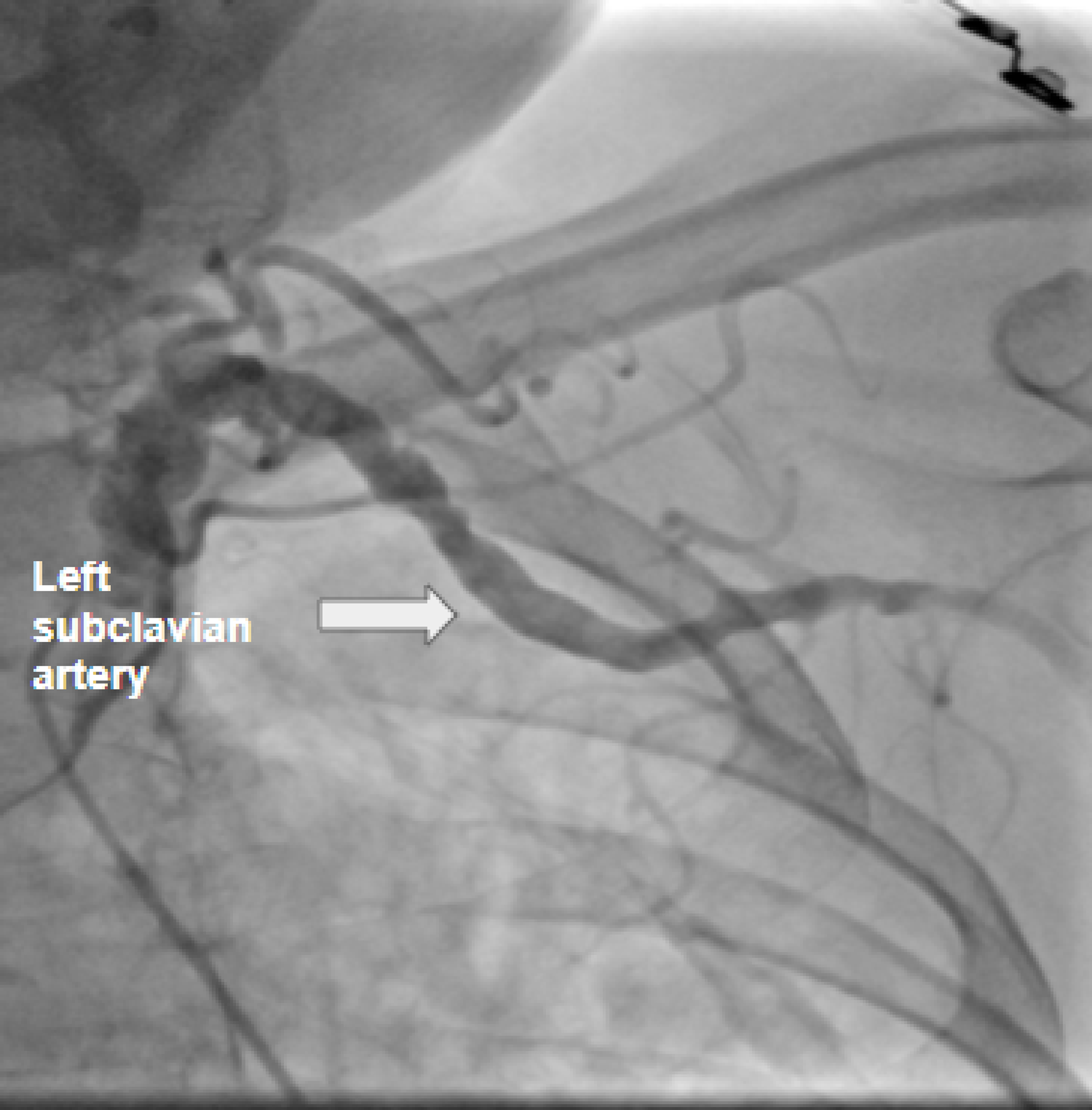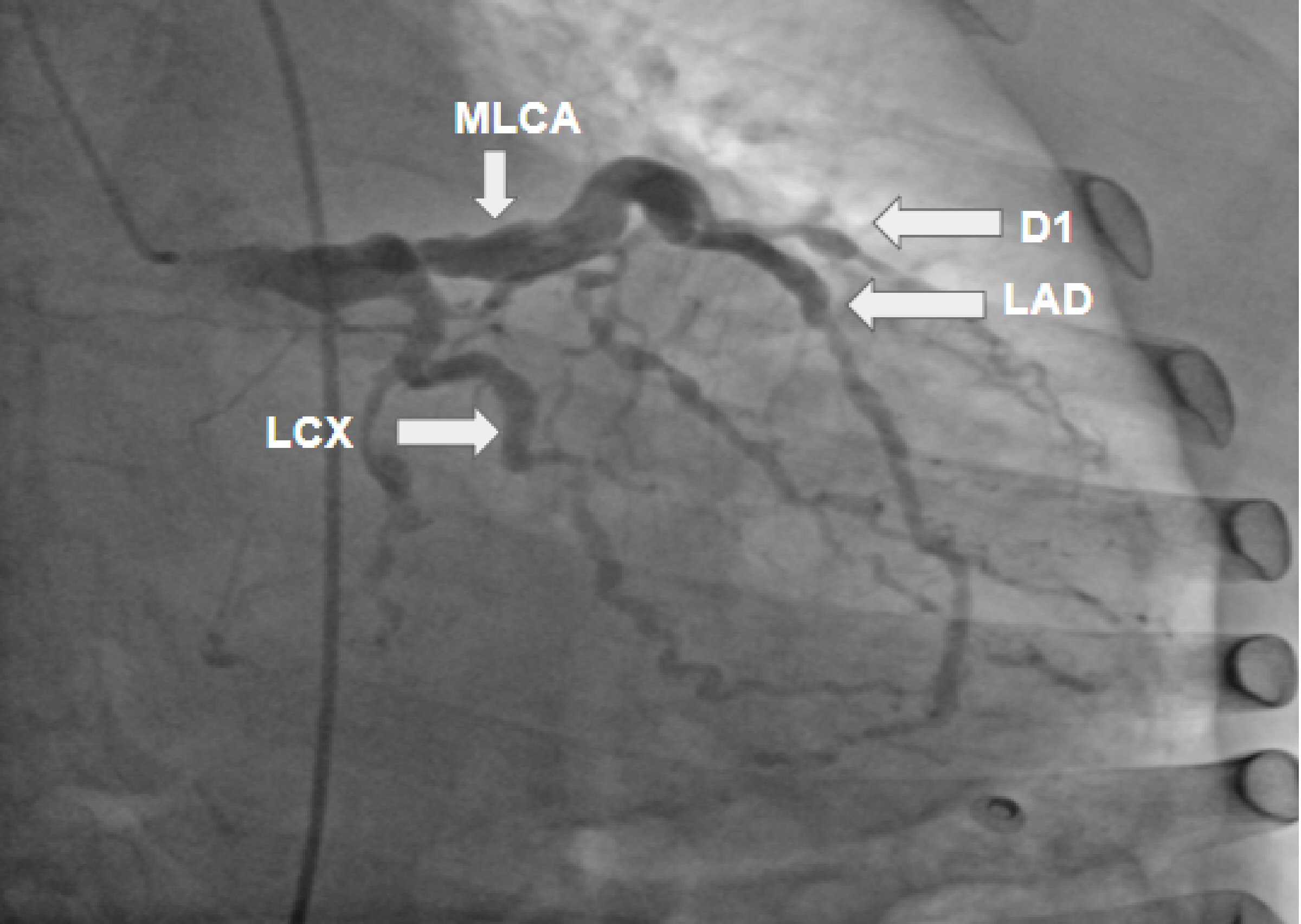
Figure 1. Coronary artery angiogram revealing a severely ectatic left main coronary artery (LMCA); an extremely ectatic left anterior descending (LAD) artery with multiple areas of moderate-to-severe distal and apical stenosis; severely ectatic and stenotic areas of diagonal 1 artery (D1); and multiple areas of stenosis including the first obtuse marginal (OM1) and distal left circumflex (LCX) arteries.
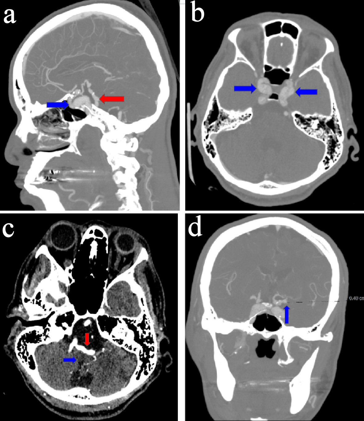
Figure 3. (a) Sagittal image of a computerized tomography (CT) angiogram of the head, revealing ectasia of the vertebrobasilar artery (red arrow) and calcification, dilatation, and ectasia of the intracavernous segment of the left internal carotid artery (blue arrow). (b) Axial image of a CT angiogram of the head, revealing bilateral dilatation and ectasia of the intracavernous segments of the internal carotid arteries (blue arrows). (c) Axial image of a CT angiogram of the head, revealing ectasia involving the vertebrobasilar artery (red arrow) and causing a mass effect on the medulla (blue arrow). (d) Coronal image of a CT angiogram of the head, revealing a 4-mm supraclinoid left internal carotid aneurysm (blue arrow).