Journal of Medical Cases, ISSN 1923-4155 print, 1923-4163 online, Open Access
Article copyright, the authors; Journal compilation copyright, J Med Cases and Elmer Press Inc
Journal website http://www.journalmc.org

Case Report

Volume 10, Number 9, September 2019, pages 284-287


A Patient With Sporotrichosis Diagnosed By Molecular Biology Combined With Traditional Methods

Figures

Figure 1.
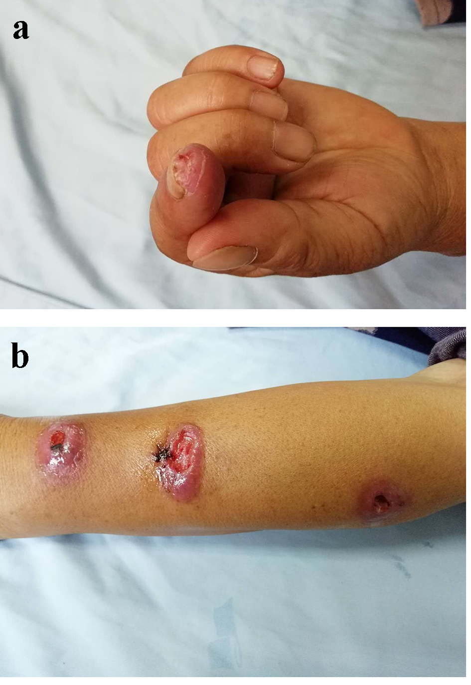
Figure 1. (a) The appearance of skin lesion at the tip of the left index finger. (b) The appearance of skin lesion on the left forearm.
Figure 2.
Figure 2. (a) The appearance of colonies cultured in the Sabouraud medium for 10 days. (b) The morphology of fungi from cultured colonies under the fluorescence through a microscope.
Figure 3.
Figure 3. (a) Histopathological images of lesion tissue. Diffuse mixed inflammatory cell infiltration in the dermis, including lymphocyte, neutrophil, eosinophil and plasma cells (H&E, × 100). (b) Histopathological images of lesion tissue and a red-stained spore in the deep dermis (PAS, × 400). H&E: hematoxylin and eosin; PAS: periodic acid-Schiff.