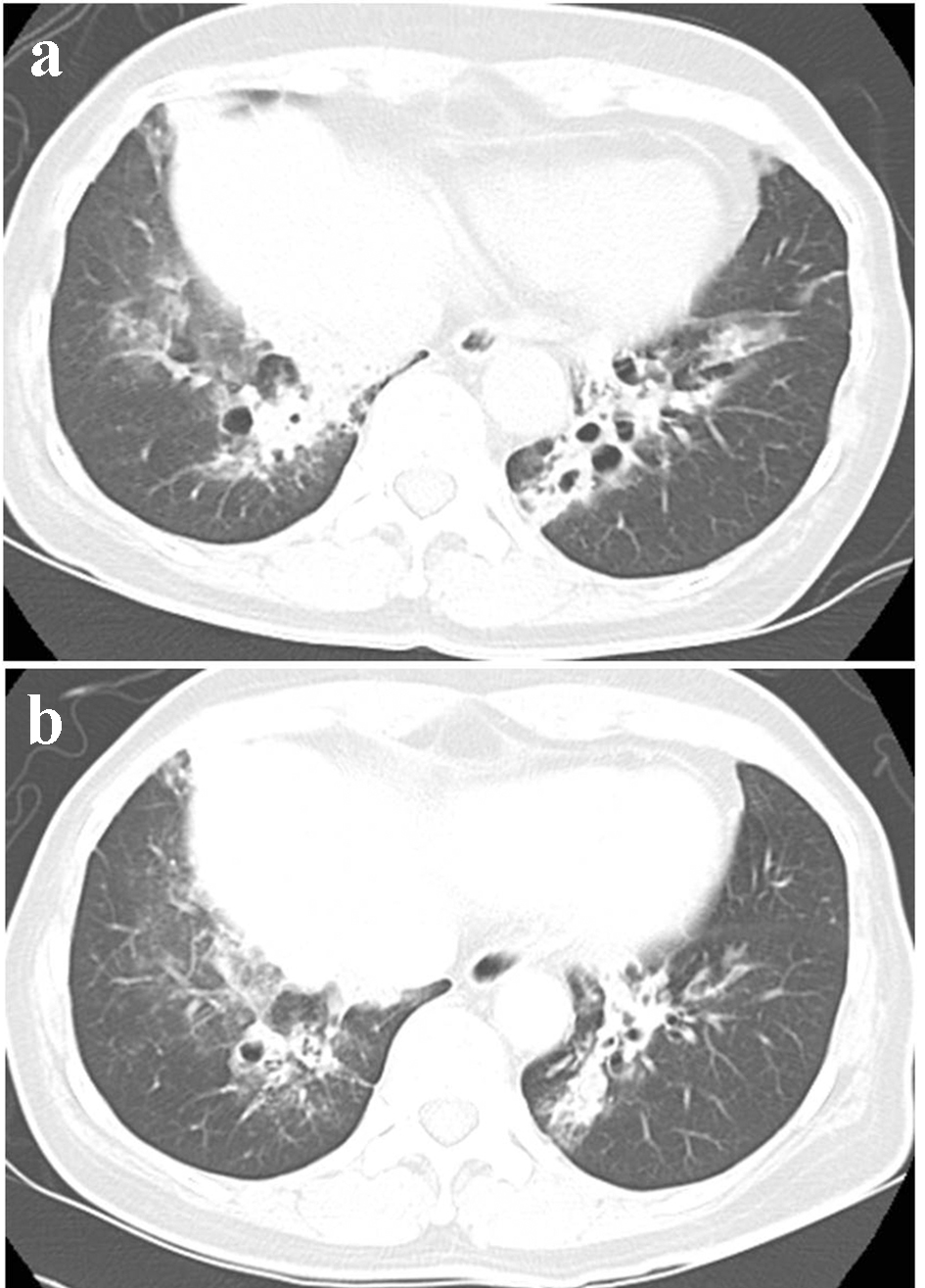
Figure 1. Bronchiectasis (a) at this admission and (b) 5 years ago.
| Journal of Medical Cases, ISSN 1923-4155 print, 1923-4163 online, Open Access |
| Article copyright, the authors; Journal compilation copyright, J Med Cases and Elmer Press Inc |
| Journal website http://www.journalmc.org |
Case Report
Volume 10, Number 9, September 2019, pages 267-270
Growth of Mediastinal Bronchial Artery Aneurysms in a Patient With Bronchiectasis
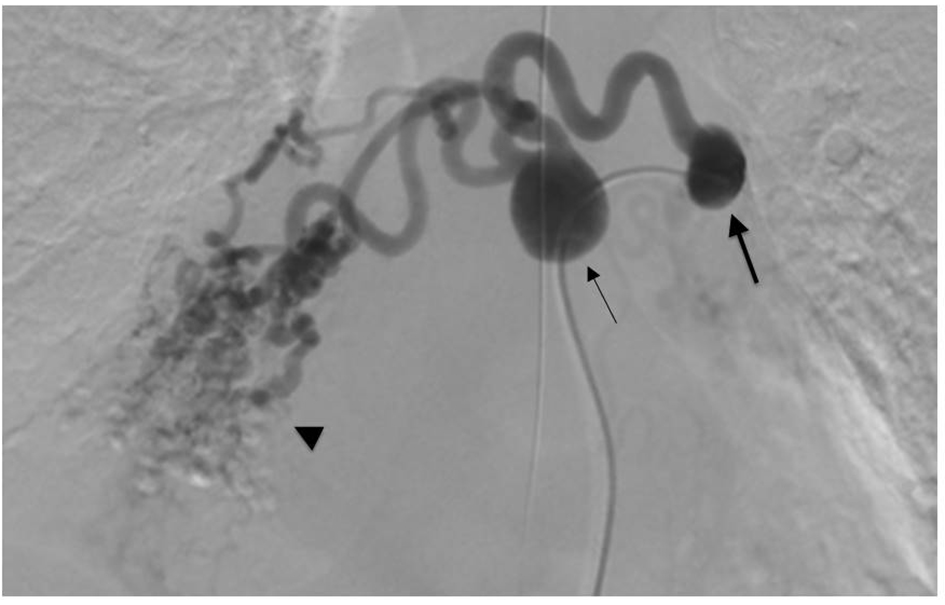
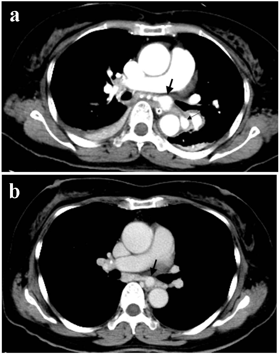
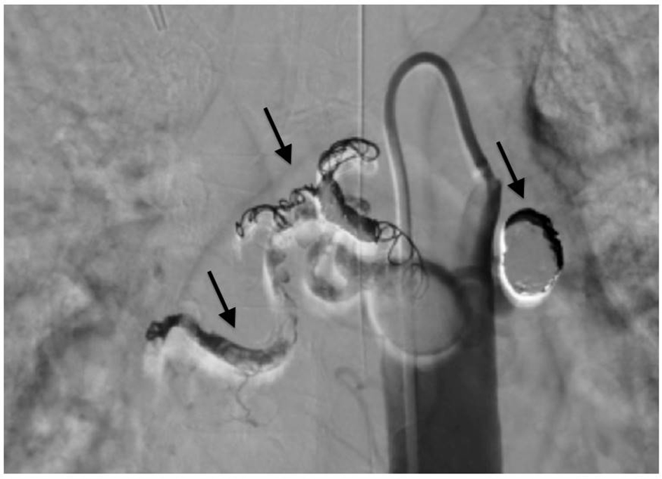
Figures