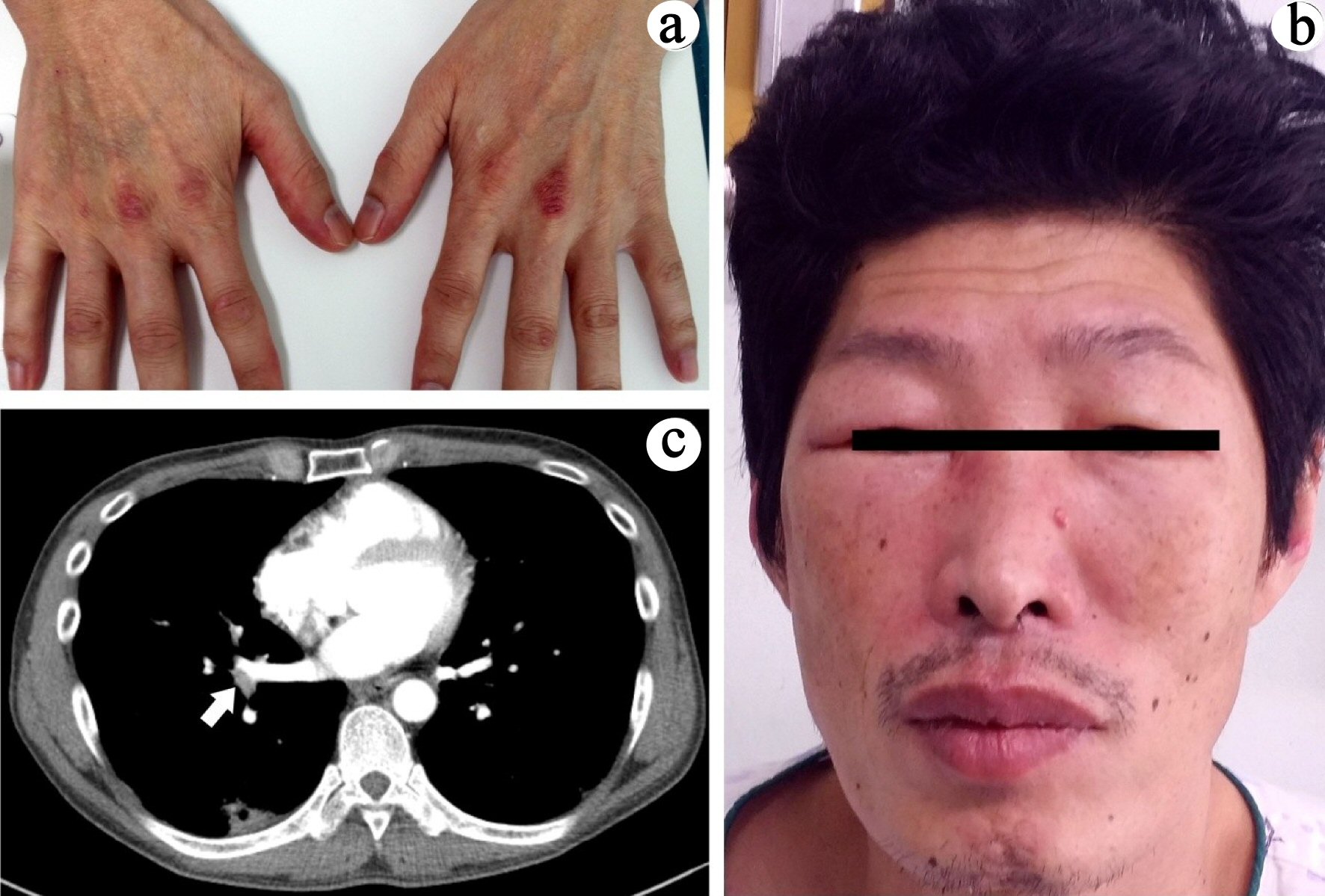
Figure 1. (a) Gottron’s papules on both hands. (b) Both periorbital edema and heliotrope rash. (c) Focal wedge-shaped consolidation in right lower lobe and filling defect in the distal right lower pulmonary vein (arrow).
| Journal of Medical Cases, ISSN 1923-4155 print, 1923-4163 online, Open Access |
| Article copyright, the authors; Journal compilation copyright, J Med Cases and Elmer Press Inc |
| Journal website http://www.journalmc.org |
Case Report
Volume 10, Number 9, September 2019, pages 280-283
Significant Muscle Hemorrhage Associated With Low-Molecular-Weight Heparin Use in Dermatomyositis: A Case Report
Figures
Table
| Case | Age | Sex | Bleeding sites | Type of anticoagulant | Onset after administration of anticoagulant | Coagulability | Treatment | Outcome | Reference |
|---|---|---|---|---|---|---|---|---|---|
| M: male; F: female; UFH: unfractionated heparin; APTT: activated partial thromboplastin time. | |||||||||
| 1 | 80 | M | Left rectus sheath, oblique, right thigh | UFH | 9 days | APTT prolonged | Transfusion | Alive | [3] |
| 2 | 77 | F | Left iliopsoas, left iliacus, retroperitoneum | UFH, ticlopidine | 4 days | APTT prolonged | Embolization | Death | [4] |
| 3 | 64 | F | Right psoas, right iliacus, retroperitoneum, left rectus sheath | Dalteparin | 9 days | Normal | Embolization | Death | [5] |
| 4 | 65 | F | Both iliopsoases, thighs | UFH | 4 days | APTT prolonged | Transfusion | Alive | [6] |
| 5 | 60 | M | Left deltoid, left trapezius | UFH | 6 days | APTT prolonged | Transfusion | Death | [7] |
| 6 | 43 | M | Left iliopsoas, left iliacus, retroperitoneum | Enoxaparin | 10 days | Normal | Transfusion | Alive | Our case |