Journal of Medical Cases, ISSN 1923-4155 print, 1923-4163 online, Open Access
Article copyright, the authors; Journal compilation copyright, J Med Cases and Elmer Press Inc
Journal website http://www.journalmc.org

Case Report

Volume 10, Number 8, August 2019, pages 253-256


Renal Metastasis of Small Cell Lung Cancer With Urothelial Carcinoma of the Bladder Misdiagnosed as Renal Cell Carcinoma

Figures

Figure 1.
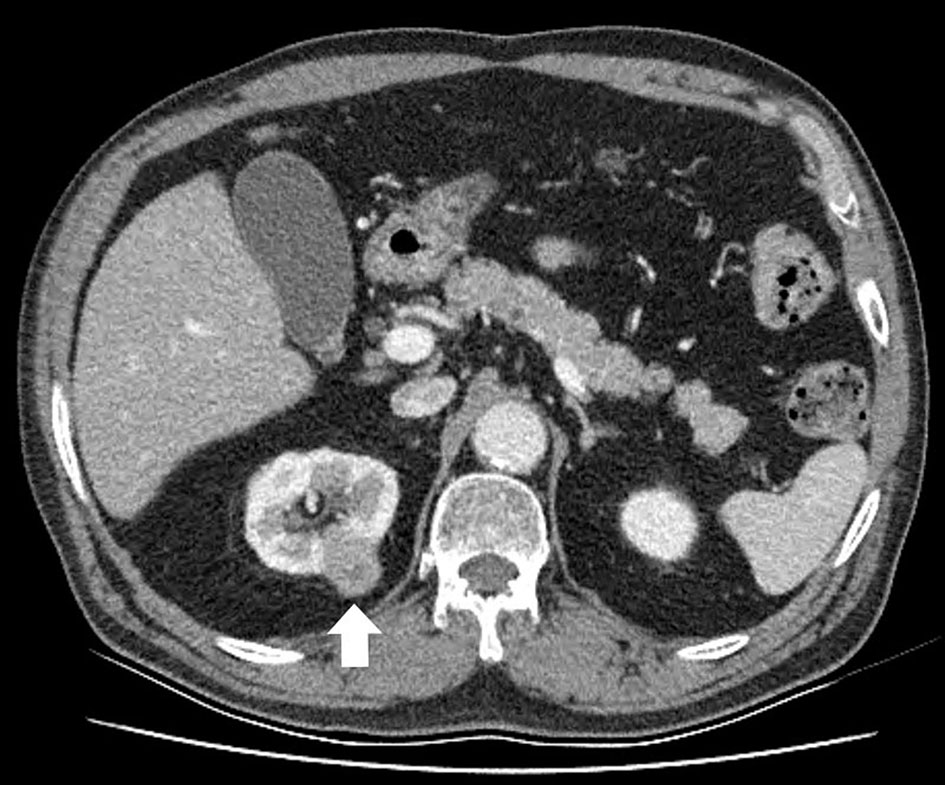
Figure 1. An abdominal computed tomography scan reveals the right renal mass. The tumor is seen as an irregular and ill-defined heterogeneous contrast-enhanced lesion.
Figure 2.
Figure 2. Gross picture of the excised renal tumor and kidney. The tumor directly invades the perirenal fat, but not beyond Gerota’s fascia (pT3a).
Figure 3.
Figure 3. Renal biopsy specimen (original magnification, × 400): hematoxylin-eosin staining (a) and immunohistochemical staining of CD56 (b), TTF-1 (c) and bladder biopsy specimen (× 400): hematoxylin-eosin staining (d).
Figure 4.
Figure 4. Maximum intensity projection (MIP) image from an positron emission tomography/computed tomography (PET/CT) scan with 18F-fluorodeoxyglucose (F18 FDG) shows intense uptake in the lung mass invading the central left upper lobe (arrow) and an oval focus of intense uptake in the right hepatic lobe (arrowhead). Post-operation change shows diffuse uptake in the right lower quadrant of the abdominal wall.