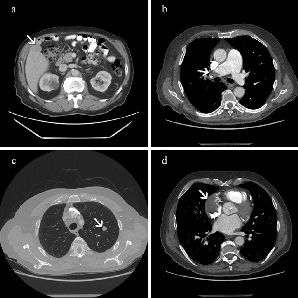
Figure 1. (a) Contrast-enhanced CT scan performed on the day of admission. The abdominal image acquired in portal venous phase shows a slightly hypodense bulging mass in the right lobe of the liver (arrow). (b) CT scan shows a large filling defect representing thrombus in the right pulmonary artery (arrow). There is an enlargement of the main pulmonary trunk and right pulmonary artery. (c) On CT scan there is a nodule measuring 1 cm in diameter in the upper lobe of the left lung (arrow). (d) CT scan shows a large filling defect in the right atrium (arrow) of the heart with subtotal obliteration of the appendage. CT: computed tomography.
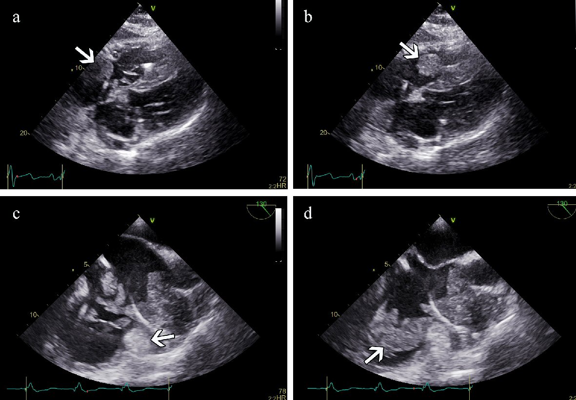
Figure 2. (a) TTE (subcostal view) in systole. A mass is detected in the right atrium (arrow). (b) TTE in diastole. The same mass is noticed in the right ventricular inflow tract (arrow). (c) TEE in systole. An immobile structure is demonstrated in the RAA (arrow) surrounding the pacemaker electrode. Mobile masses are demonstrated in the RA. (d) TEE in diastole. Mobile masses have moved into the RV inflow tract (arrow). TTE: transthoracic echocardiogram; TEE: transesophageal echocardiography; RAA: right atrial appendage.

Figure 3. (a) Primary cardiac extraskeletal osteosarcoma. Pleomorphic tumor cells (arrow) and normal myocardium (H&E, × 20). (b) Osteoclast-like giant cells (arrow) (H&E, × 20). (c) Neoplastic osteoid matrix (arrow) (H&E, × 20). (d) Giant and spindle cells (arrow) (H&E, × 40).
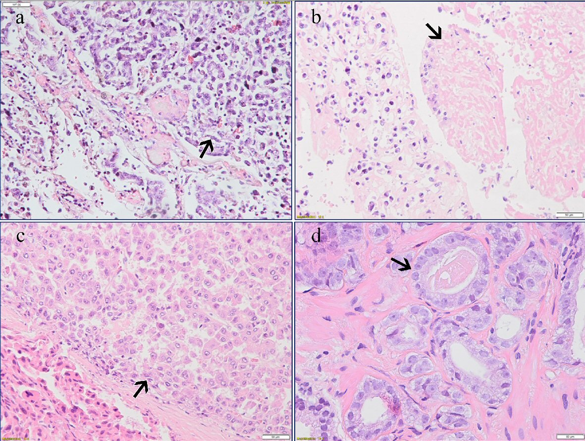
Figure 4. (a) Metastatic pulmonary osteosarcoma (arrow) (H&E, × 20). (b) Tumor thrombus in the lung (arrow) (H&E, × 20). (c) Hepatocellular carcinoma (arrow) (H&E, × 20). (d) Acinar adenocarcinoma of the prostate (arrow) (H&E, × 40).