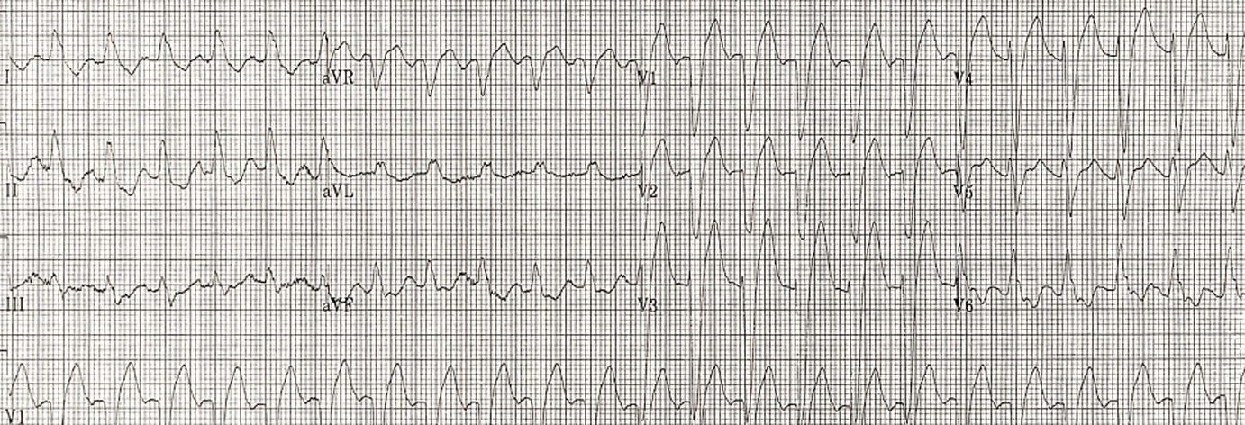
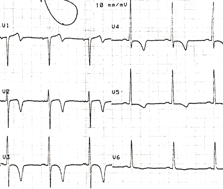
Figure 1. ECG initial presentation.
| Journal of Medical Cases, ISSN 1923-4155 print, 1923-4163 online, Open Access |
| Article copyright, the authors; Journal compilation copyright, J Med Cases and Elmer Press Inc |
| Journal website http://www.journalmc.org |
Case Report
Volume 10, Number 7, July 2019, pages 193-197
Wellens’ Syndrome: An Atypical Localization After an Atypical Transient Left Bundle Branch Block Presentation
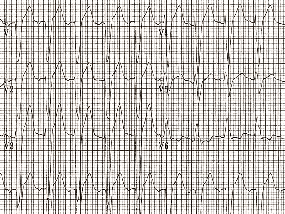
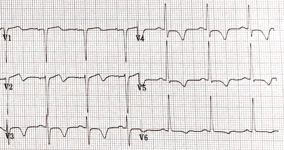
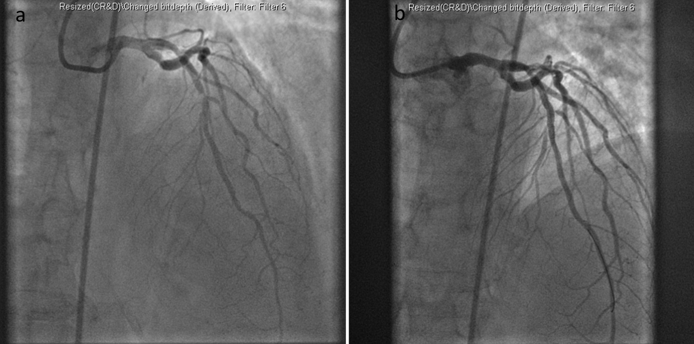
Figures
Table
| Criteria for Wellens’ syndrome |
|---|
| Wellens’ type A or type B wave(s) in leads V2 and V3, occasionally in leads V1-V6 |
| Recent history of angina |
| Pattern present during pain-free state |
| Isoelectric or minimally elevated (< 1 mm) ST segment |
| No precordial Q waves and preserved R wave progression |
| Normal or slightly elevated cardiac serum markers |