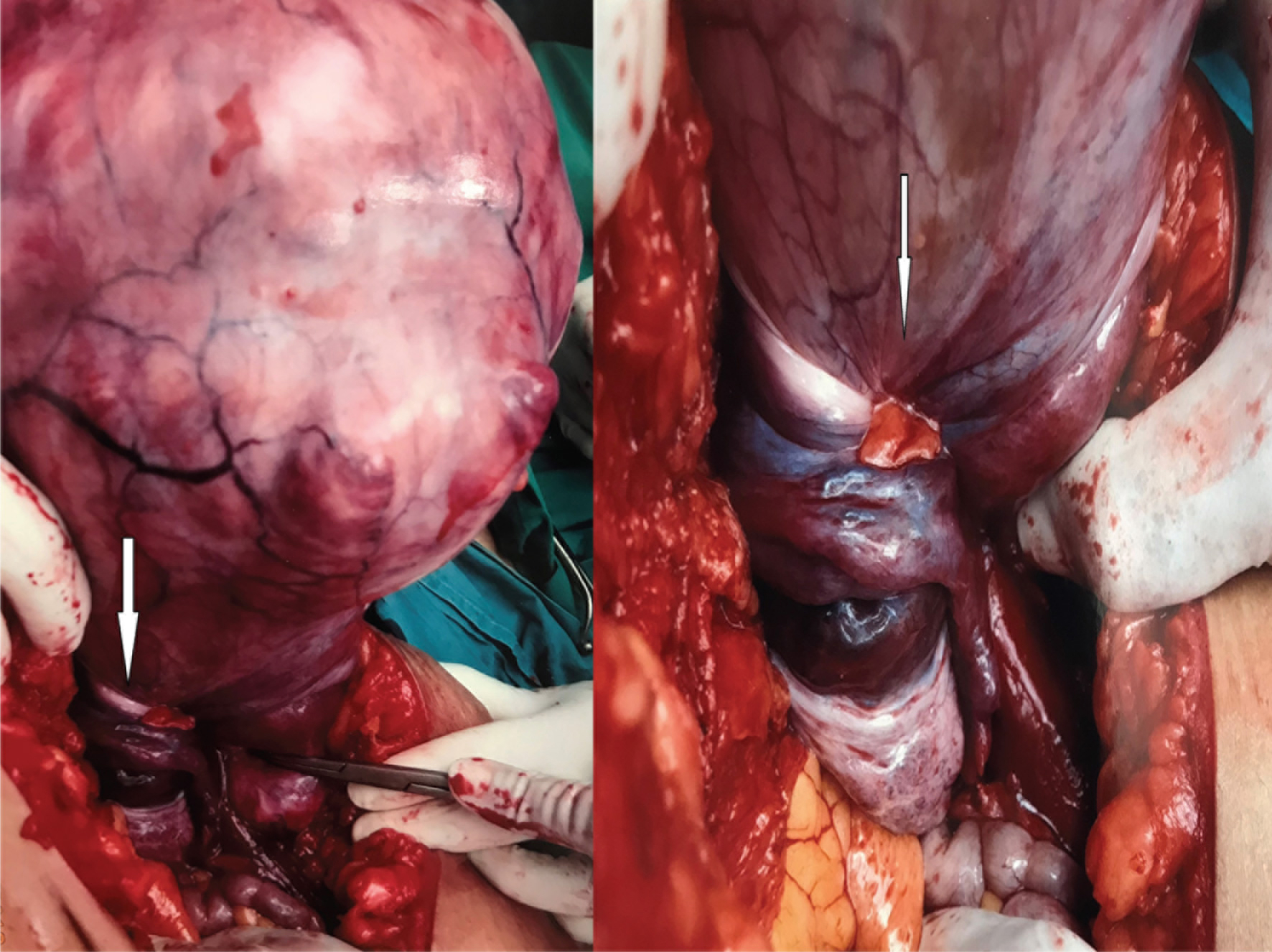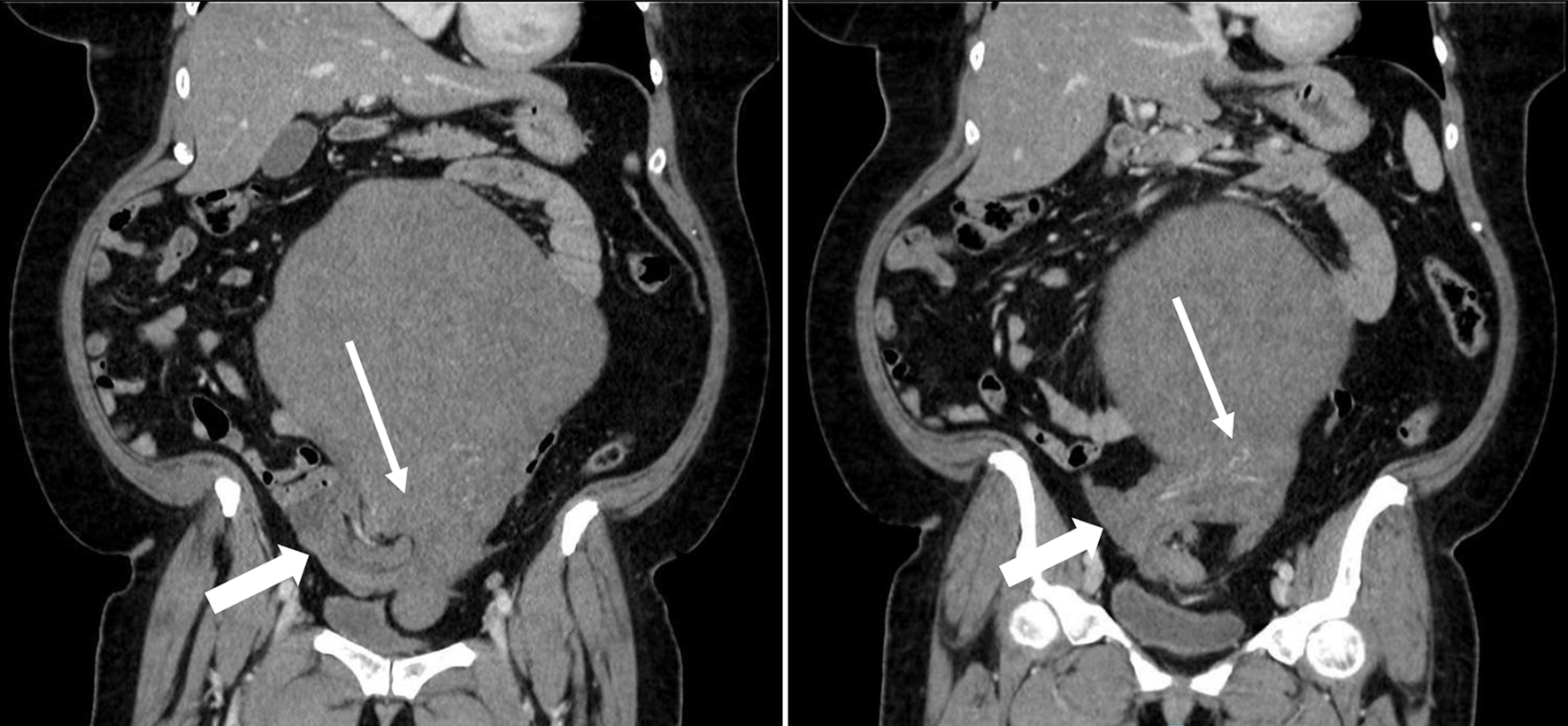
Figure 1. Contrast-enhanced CT of abdomen and pelvis (coronal section) showed torsion of lower uterine segment (white arrow) and bilateral adnexa at the right hemipelvis (blocked white arrow).
| Journal of Medical Cases, ISSN 1923-4155 print, 1923-4163 online, Open Access |
| Article copyright, the authors; Journal compilation copyright, J Med Cases and Elmer Press Inc |
| Journal website http://www.journalmc.org |
Case Report
Volume 10, Number 6, June 2019, pages 164-167
A Rare Case of Complete Uterine Torsion in a Postmenopausal Woman
Figures