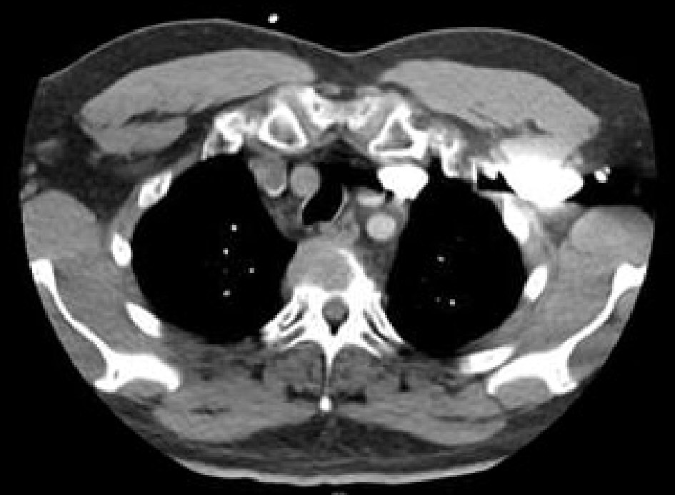
Figure 1. A large mediastinal mass with multiple hypodensities.
| Journal of Medical Cases, ISSN 1923-4155 print, 1923-4163 online, Open Access |
| Article copyright, the authors; Journal compilation copyright, J Med Cases and Elmer Press Inc |
| Journal website http://www.journalmc.org |
Case Report
Volume 10, Number 2, February 2019, pages 37-40
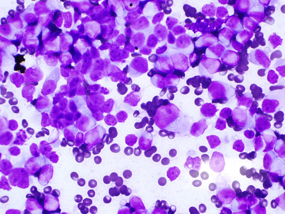
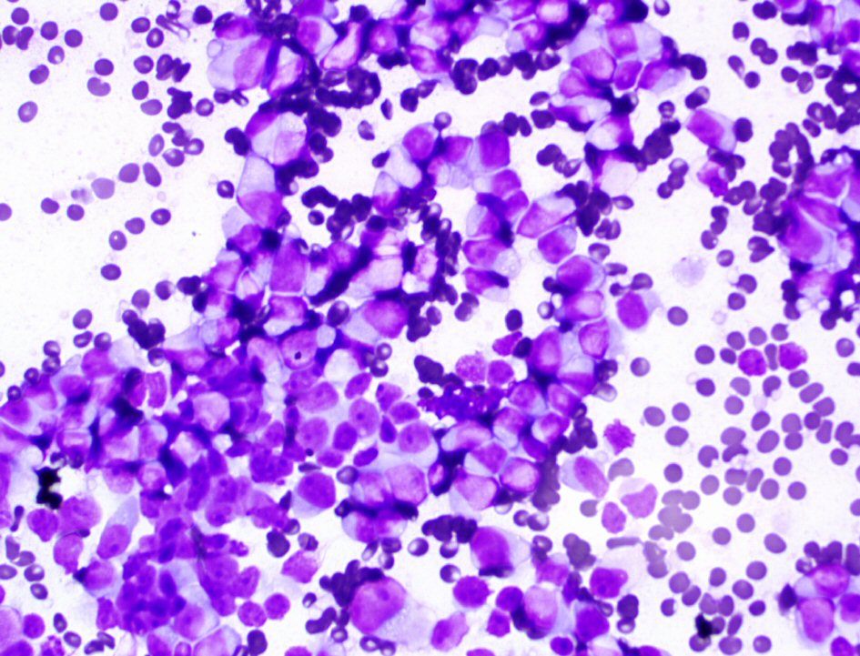
Utility of Fine Needle Aspiration for Diagnosis of Plasmacytoma
Figures
Table
| NO. | EMP locations | Country | Authors |
|---|---|---|---|
| 1 | Gall Bladder | USA | St. Romain et al [4] |
| 2 | Liver | Germany | Bangerter et al [1] |
| 3 | Tonsil | India | Bhat et al [6] |
| 4 | Anterior Mediastinum | USA, Japan, Taiwan | Mallo et al [12] |
| 5 | Larynx | USA | Saad et al [9] |