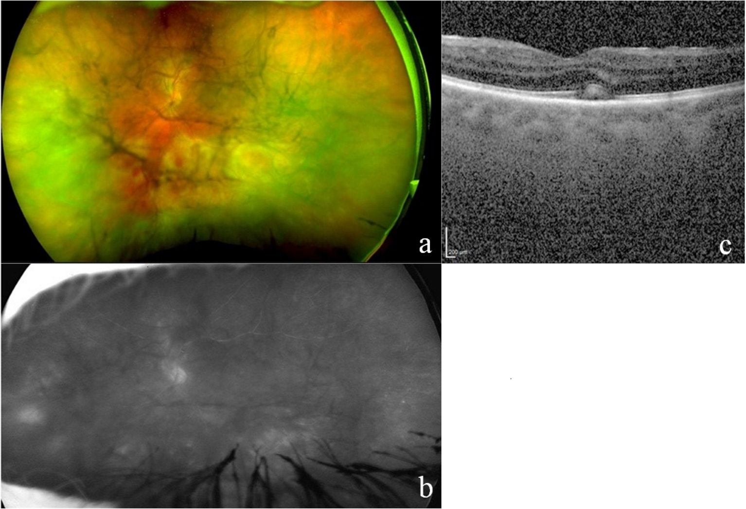
Figure 1. (a) Color fundus photograph with hemorrhage and yellowish subretinal infiltrates present up to the inferior arcade in the left eye with dense vitritis and vitreous haze. (b) Fluorescein angiography demonstrating pronounced leakage from disc and inferior vessels in the left eye despite a hazy view of the fundus. (c) Optical coherence tomography revealing indistinct choroidal details, foveal outer retinal band elevation and disruption in the left eye, with a hyperintense subretinal infiltrate in addition to a mild epiretinal membrane.
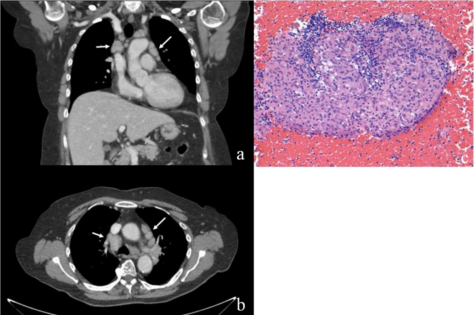
Figure 2. (a, b) Coronal and axial chest CT with contrast demonstrating bilateral hilar lymphadenopathy (white arrows). (c) Low magnification (× 200) H&E stain of hilar biopsy showing extensive granulomatous inflammation with no caseation.
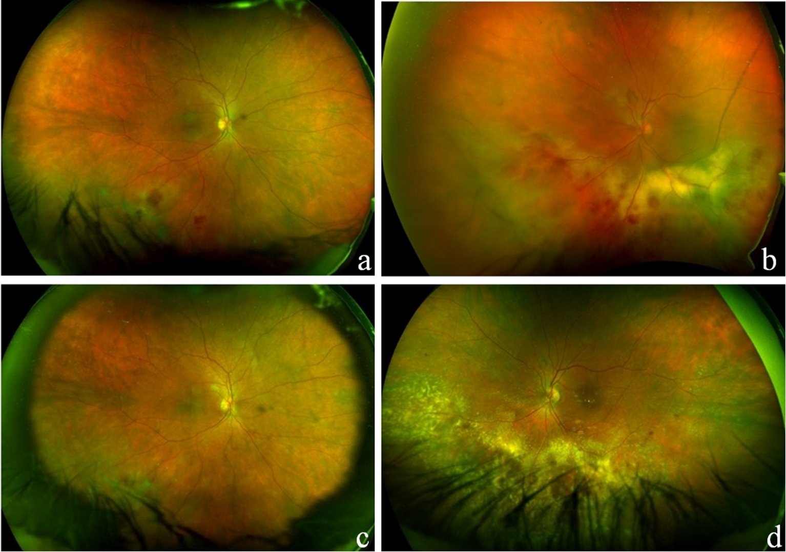
Figure 3. Fundus examination following left vitrectomy/vitreous biopsy before initiation of treatment (a, b) and after 4 months of oral prednisone treatment (c, d). After treatment photograph demonstrates retinal exudates and mild hemorrhage (left more than right) and no significant vitreous haze in either eye.