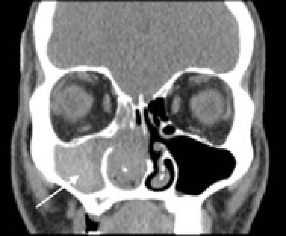
Figure 1. CT showing right nasal sinus opacification (see arrow).
| Journal of Medical Cases, ISSN 1923-4155 print, 1923-4163 online, Open Access |
| Article copyright, the authors; Journal compilation copyright, J Med Cases and Elmer Press Inc |
| Journal website http://www.journalmc.org |
Case Report
Volume 9, Number 11, November 2018, pages 371-374
Localized Extramedullary Plasmacytoma: A Report of Two Cases
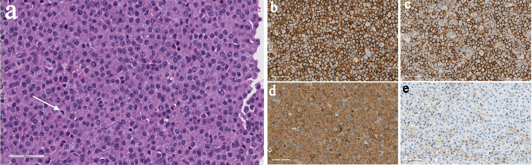
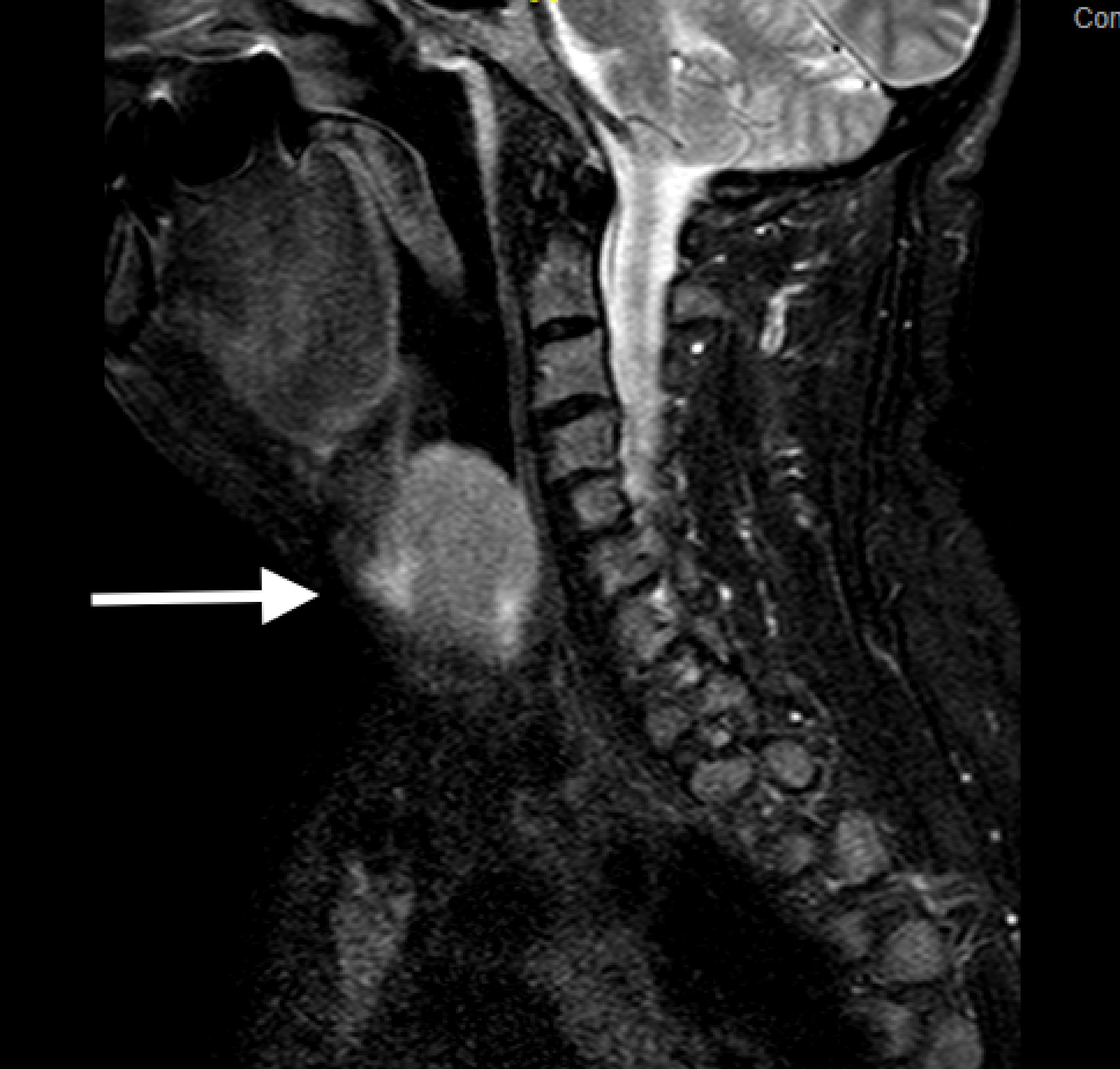
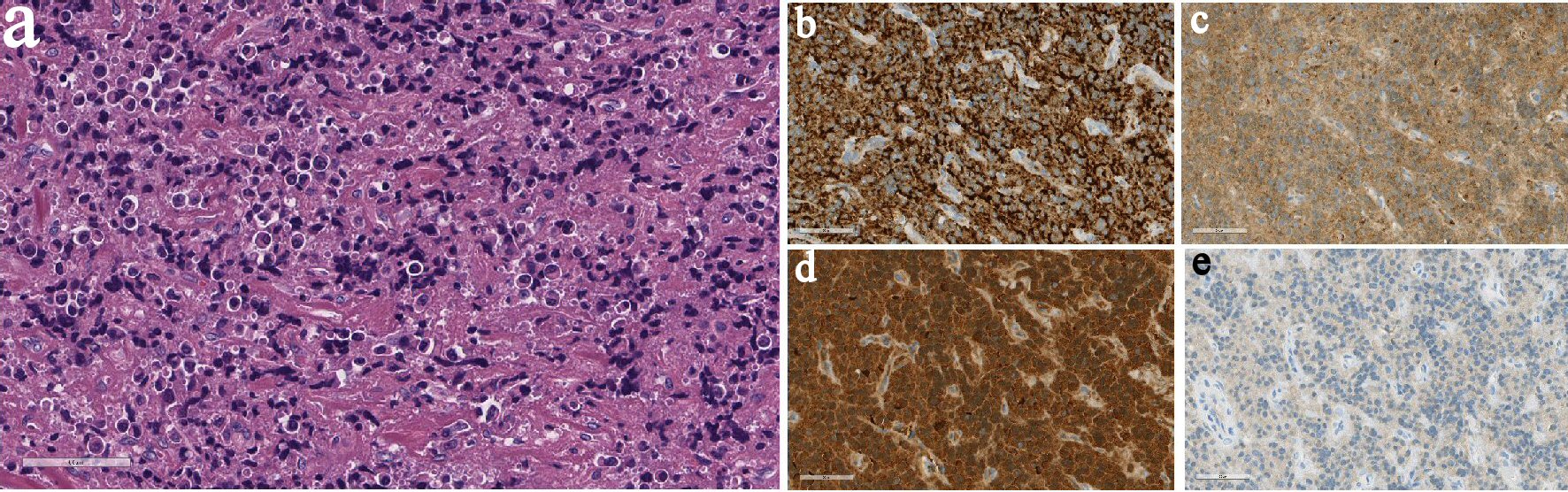
Figures