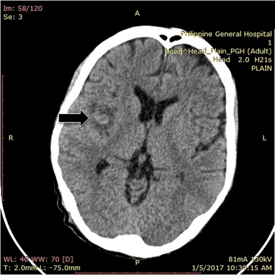
Figure 1. Plain cranial CT scan showing right middle cerebral infarct with hemorrhagic conversion.
| Journal of Medical Cases, ISSN 1923-4155 print, 1923-4163 online, Open Access |
| Article copyright, the authors; Journal compilation copyright, J Med Cases and Elmer Press Inc |
| Journal website http://www.journalmc.org |
Case Report
Volume 9, Number 8, August 2018, pages 246-251
“Cardiovascular Shower” Infective Endocarditis Causing Multiple Septic Emboli: A Case Report and a Review of Related Literature
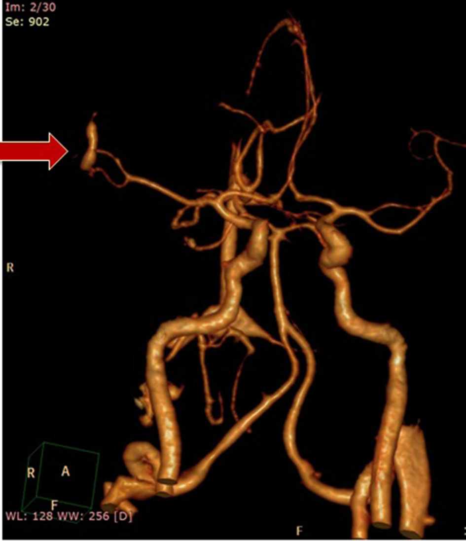
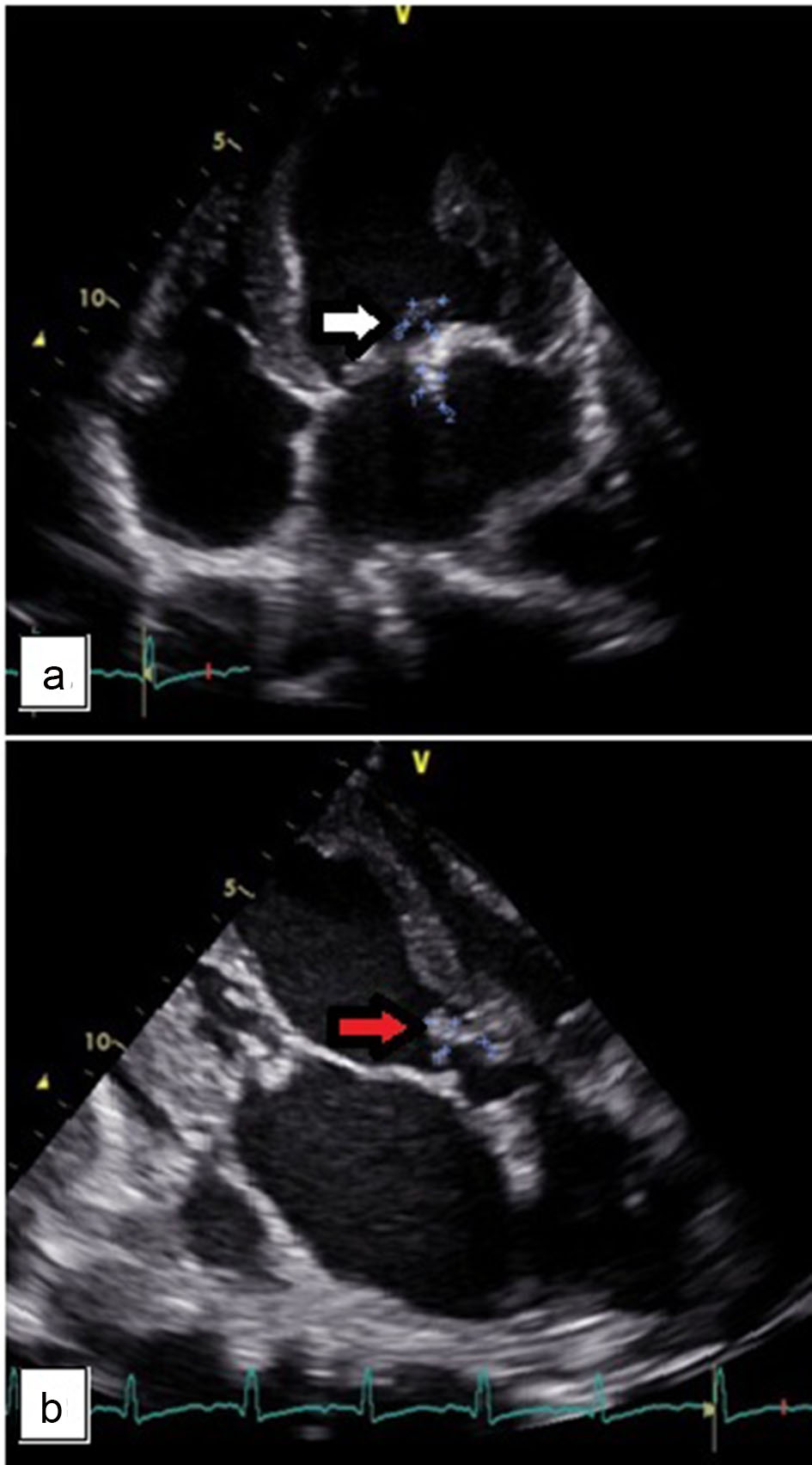
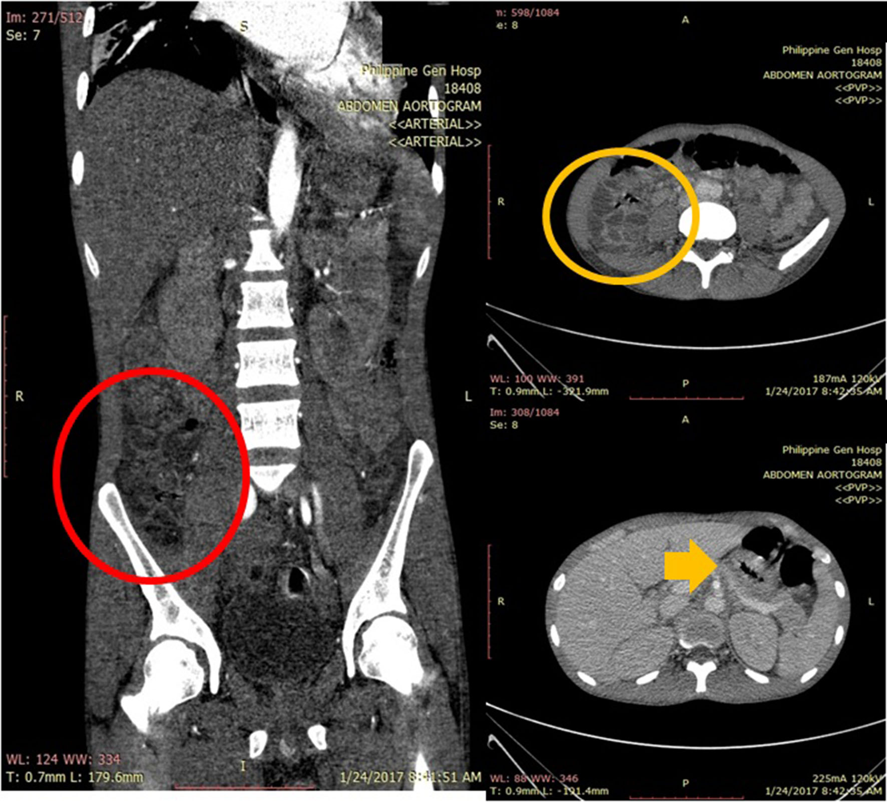
Figures