Journal of Medical Cases, ISSN 1923-4155 print, 1923-4163 online, Open Access
Article copyright, the authors; Journal compilation copyright, J Med Cases and Elmer Press Inc
Journal website http://www.journalmc.org

Case Report

Volume 9, Number 7, July 2018, pages 211-214


An Unusual Case of Constrictive Pericarditis in a Young Patient With Childhood History of Successfully Treated Kawasaki Disease

Figures

Figure 1.
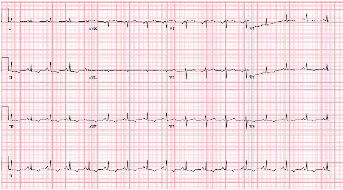
Figure 1. Electrocardiogram showing sinus rhythm, low voltage and inferolateral T wave inversion.
Figure 2.
Figure 2. (a) Parasternal long axis view of transthoracic echocardiogram (TTE) showing thickened pericardium in the inferolateral wall. (b) Showing variation on mitral inflow with respiration on TTE. (c) 2D directed M-mode on TTE showing dilated non-collapsing inferior vena cava.
Figure 3.
Figure 3. (a) Apical 4-chamber view of TTE showing thickened pericardium. (b) Pulsed wave mitral inflow on TTE showing restrictive filling pattern with E/A of 2.5. (c) Pulsed wave Doppler of mitral inflow on TTE showing variation of inflow with Valsalva maneuver.
Figure 4.
Figure 4. CT chest without contrast showing large bilateral pleural effusions associated with some compressive atelectasis. There appears to be mild diffuse pericardial thickening as opposed to hyperdense pericardial fluid.