Journal of Medical Cases, ISSN 1923-4155 print, 1923-4163 online, Open Access
Article copyright, the authors; Journal compilation copyright, J Med Cases and Elmer Press Inc
Journal website http://www.journalmc.org

Case Report

Volume 9, Number 10, October 2018, pages 328-331


Experience of Palliative Radiotherapy for Tumor-Associated Bleeding From Biliary Tract Cancer

Figures

Figure 1.
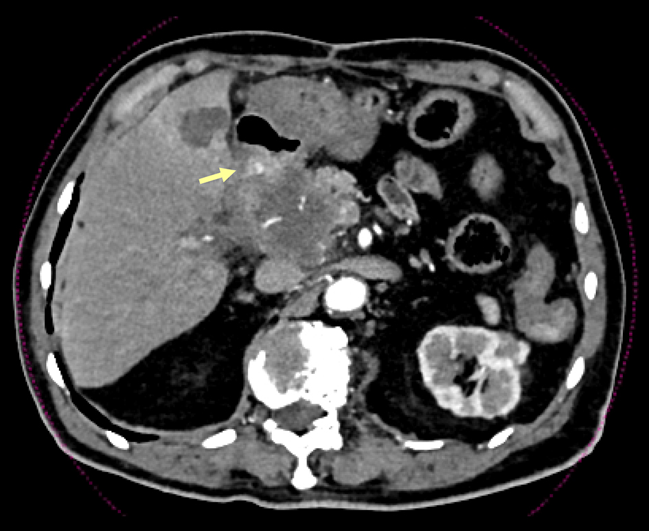
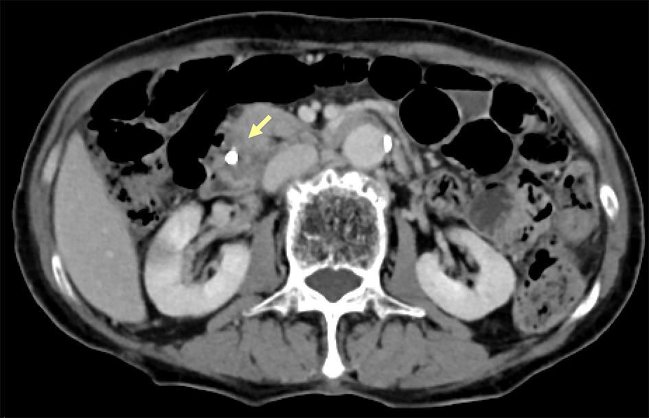
Figure 1. CT demonstrated a poorly-enhanced, low-density mass in the lower common bile duct, which invaded the duodenal wall (arrow).
Figure 2.
Figure 2. Duodenoscopy revealed sustained oozing hemorrhaging from the papilla of invasive cholangiocarcinoma.
Figure 3.
Figure 3. The clinical course of case 1.
Figure 4.
Figure 4. CT revealed both a poorly-enhanced, low-density recurrent mass at the porta hepatis, which invaded the duodenal wall, and an aneurismal dilation of the invaded-tumor vessel (arrow).
Figure 5.
Figure 5. Duodenocsopy showed a red-colored, easily-hemorrhaged ulcer with submucosal growth appearance at the supraduodenal angle.
Figure 6.
Figure 6. The clinical course of case 2.