Journal of Medical Cases, ISSN 1923-4155 print, 1923-4163 online, Open Access
Article copyright, the authors; Journal compilation copyright, J Med Cases and Elmer Press Inc
Journal website http://www.journalmc.org

Case Report

Volume 2, Number 6, December 2011, pages 262-264


Esophageal Stent Migration Into Trachea

Figures

Figure 1.
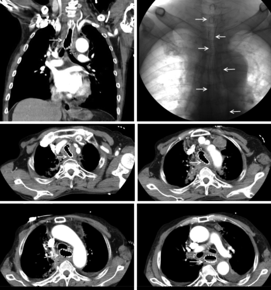
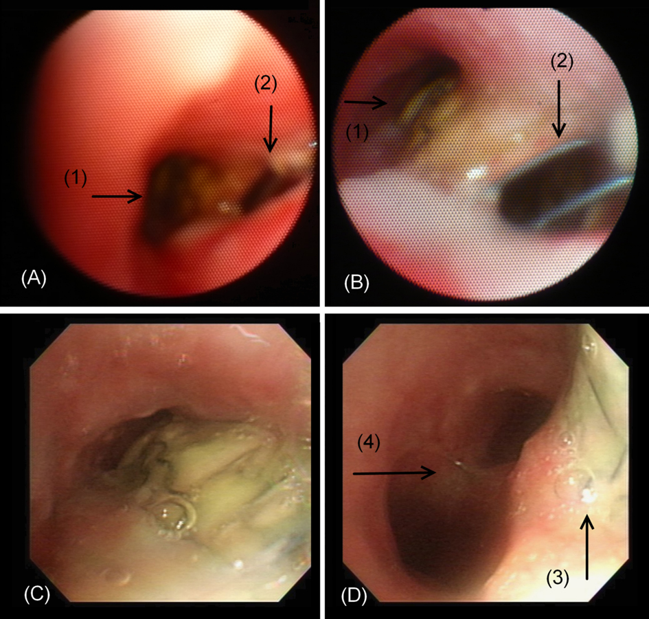
Figure 1. Bronchoscopy findings. (A) The bifurcation of tracheal lumen (1) and the TE fistula (2) mimicking the carina. (B) Close-up view of the tracheal lumen (1) showed the stent exposed through the TE fistula (2). (C) Advancing along the stenotic trachea lumen revealed the tracheal mucosal defect with stent exposure. (D) The tracheal mucosal defect with stent exposure (3) and the true carina (4).
Figure 2.
Figure 2. Computed tomography and fluoroscopy images (Solid arrows show the esophageal stent. Dotted double arrows indicate the airway, including the trachea and main bronchus).