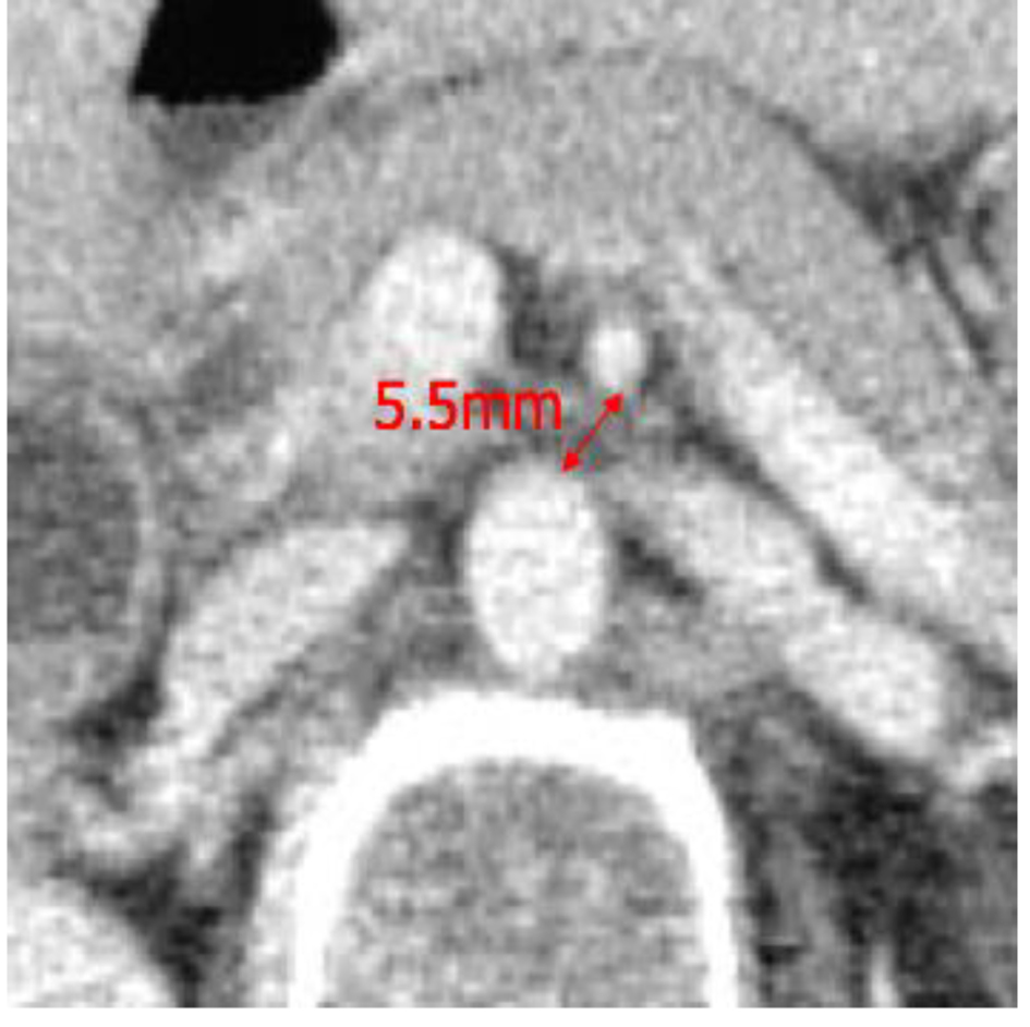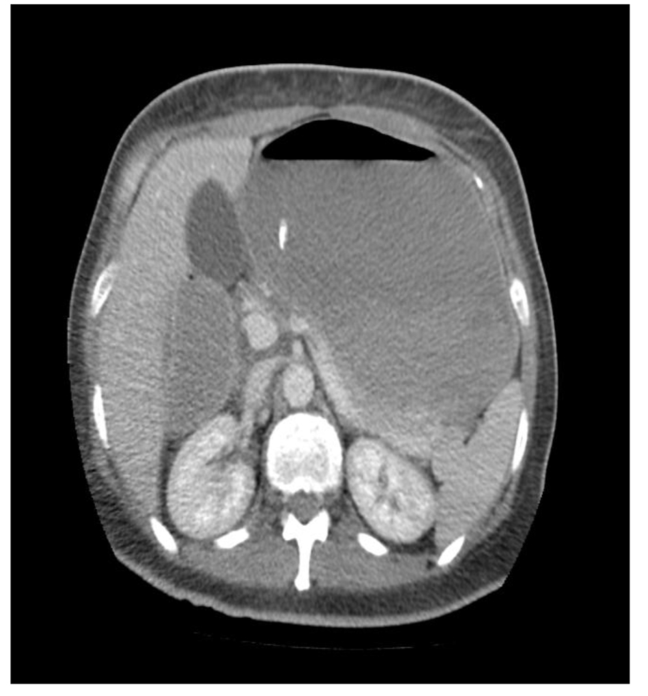
Figure 1. CT scan of abdomen showing dilatation of stomach, first and second part of duodenum. There was compression of D3 (third part of duodenum) between the aorta and SMA.
| Journal of Medical Cases, ISSN 1923-4155 print, 1923-4163 online, Open Access |
| Article copyright, the authors; Journal compilation copyright, J Med Cases and Elmer Press Inc |
| Journal website http://www.journalmc.org |
Case Report
Volume 9, Number 2, February 2018, pages 37-40
Superior Mesenteric Artery Syndrome: A High Index of Suspicion
Figures