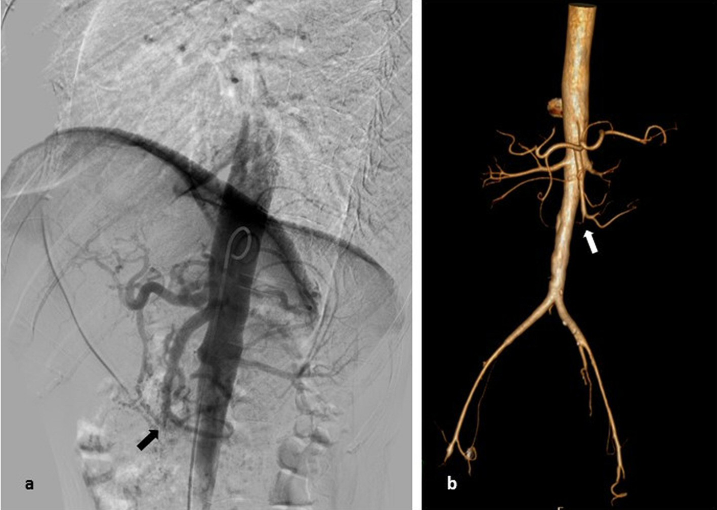
Figure 1. (a) Abdominal aortography: occlusion in the distal third of the superior mesenteric artery without collateral vascularization. (b) Abdominal CT angiography with 3D reconstruction: distal third occlusion of the superior mesenteric artery.
| Journal of Medical Cases, ISSN 1923-4155 print, 1923-4163 online, Open Access |
| Article copyright, the authors; Journal compilation copyright, J Med Cases and Elmer Press Inc |
| Journal website http://www.journalmc.org |
Case Report
Volume 8, Number 12, December 2017, pages 383-387
Severe Takayasu Arteritis Complicated by Mesenteric Ischemia
Figures