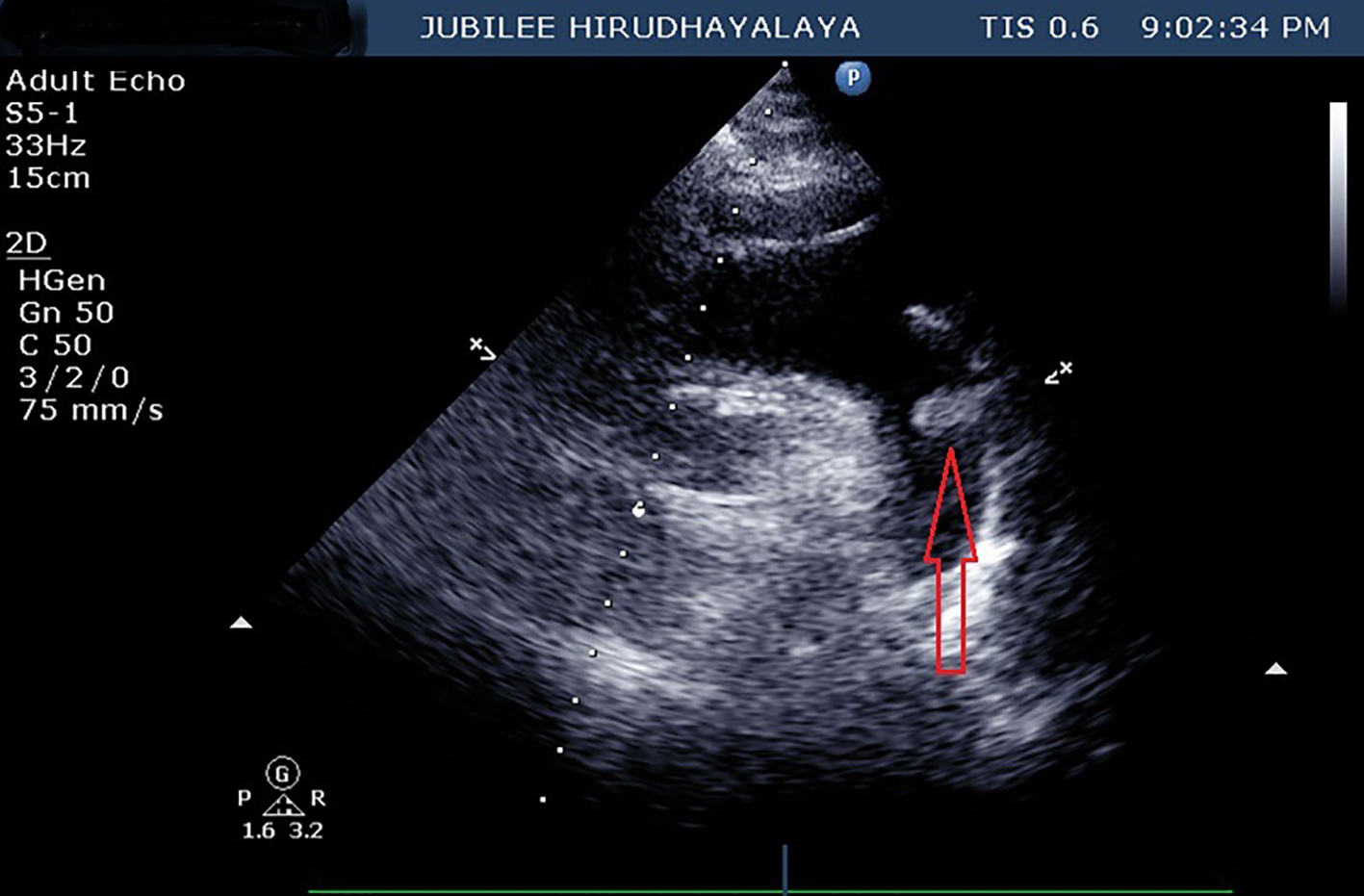
Figure 1. Parasternal long axis view on 2D echo showing thrombus (red arrow) at the level of aortic sinus.
| Journal of Medical Cases, ISSN 1923-4155 print, 1923-4163 online, Open Access |
| Article copyright, the authors; Journal compilation copyright, J Med Cases and Elmer Press Inc |
| Journal website http://www.journalmc.org |
Case Report
Volume 8, Number 11, November 2017, pages 368-370
Vanishing Aortic Thrombus
Figures