Figures
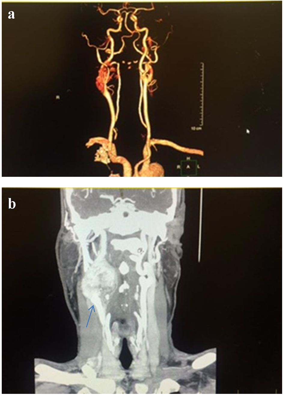
Figure 1. Computed tomography angiography (CTA) of the neck, coronal section, shows 4.9 × 3.4 × 4.5 cm enhancing mass at the carotid artery bifurcation (a) with mass effect on the right internal jugular vein and right airway (b). The carotid arteries are displaced as well.
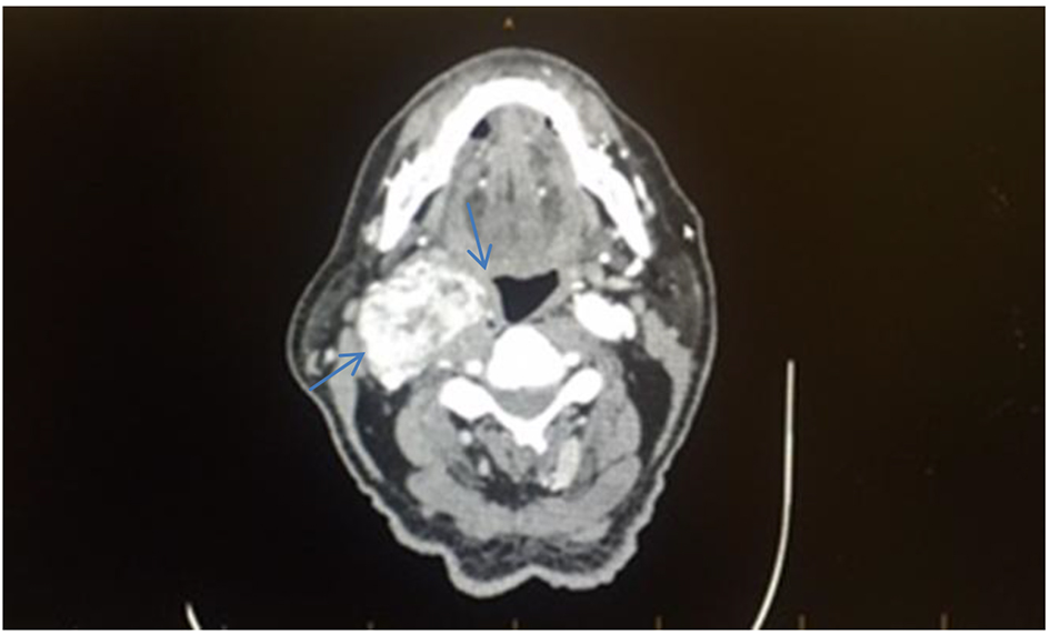
Figure 2. Computed tomography angiography (CTA) of the neck with and without contrast, axial section, shows an enhancing mass in the right side of the neck with mass effect on the right airway (blue arrows).
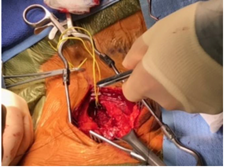
Figure 3. Perioperative finding of right side tumor that surrounds the carotid arteries.
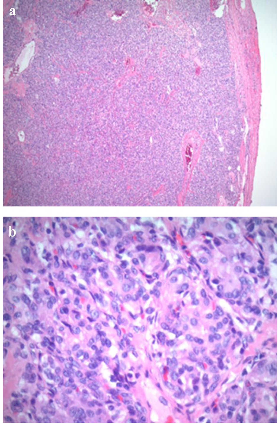
Figure 4. Low power (a: × 4) and high power light microscopy (b: × 40) showing oval cells arranged in compact cell nests (the so-called Zellballen appearance).
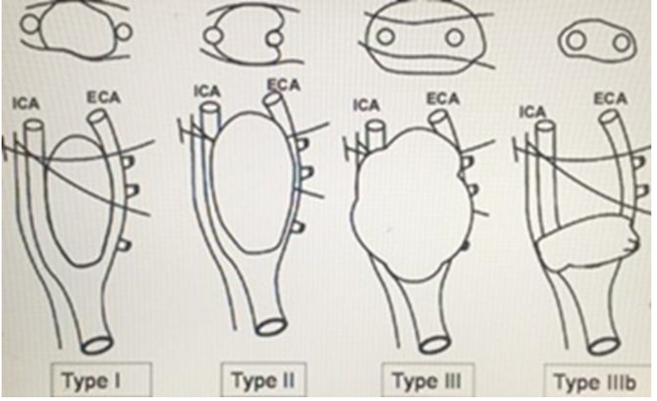
Figure 5. Schematic diagram of the Shamblin grouping of CBTs into I, II, III and IIIb. It is basically based on the relationship between the tumor and the carotid arteries. The intersected lines are the X and XII nerves which are closely related to the tumor. This diagram was adopted from Reference [18].