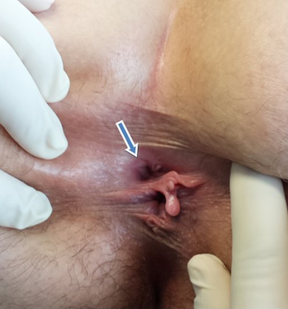
Figure 1. Visual inspection of the anus. The arrow points to the structure in the midline, posterior to the native anus.
| Journal of Medical Cases, ISSN 1923-4155 print, 1923-4163 online, Open Access |
| Article copyright, the authors; Journal compilation copyright, J Med Cases and Elmer Press Inc |
| Journal website http://www.journalmc.org |
Case Report
Volume 9, Number 2, February 2018, pages 61-63
Anal Canal Duplication in a 40-Year-Old Adult
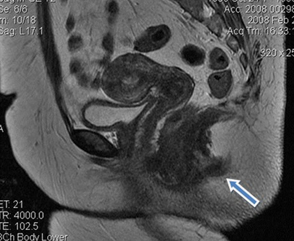
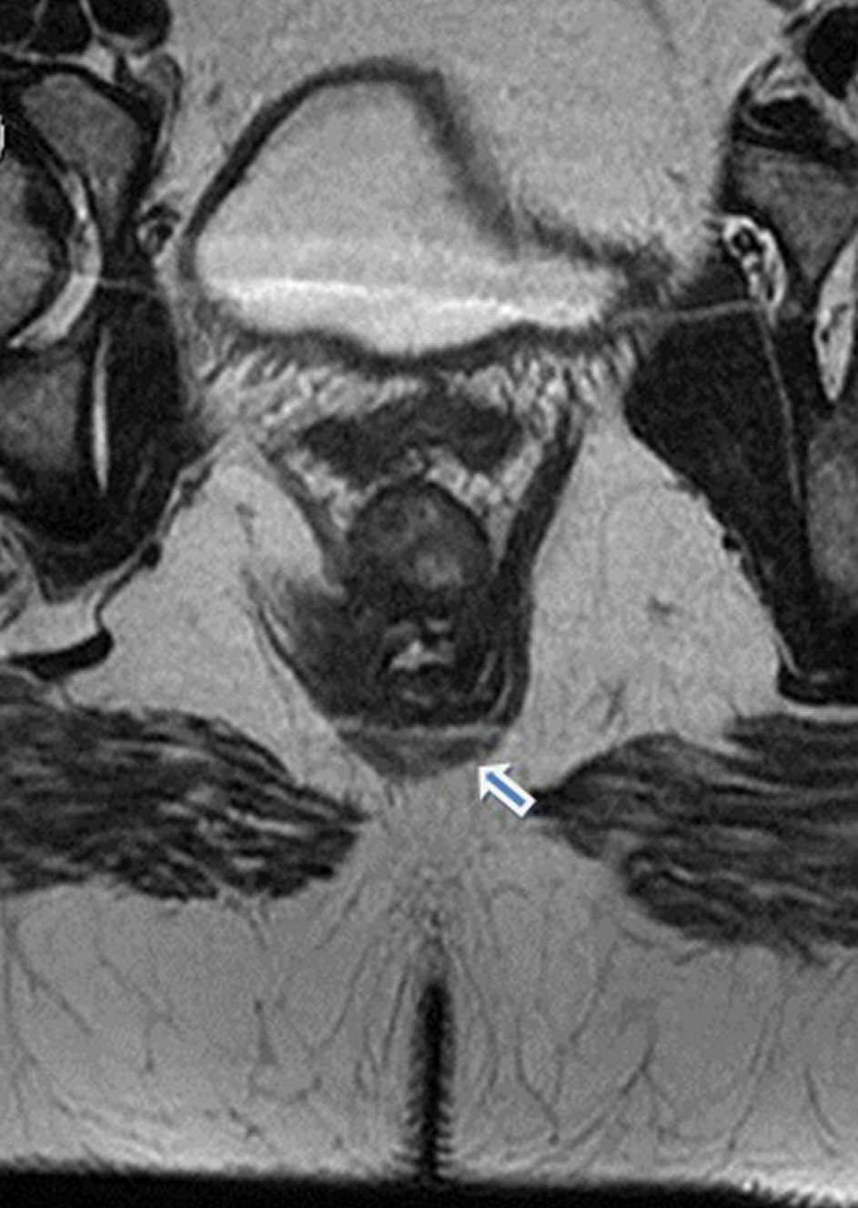
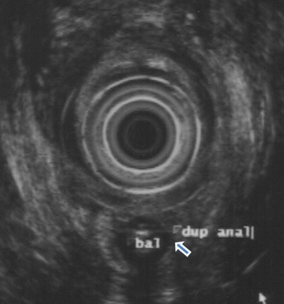
Figures