Journal of Medical Cases, ISSN 1923-4155 print, 1923-4163 online, Open Access
Article copyright, the authors; Journal compilation copyright, J Med Cases and Elmer Press Inc
Journal website http://www.journalmc.org

Case Report

Volume 8, Number 9, September 2017, pages 287-292


A Multidisciplinary Approach to a Complicated Acute Paradoxical Embolism via a Patent Foramen Ovale

Figures

Figure 1.
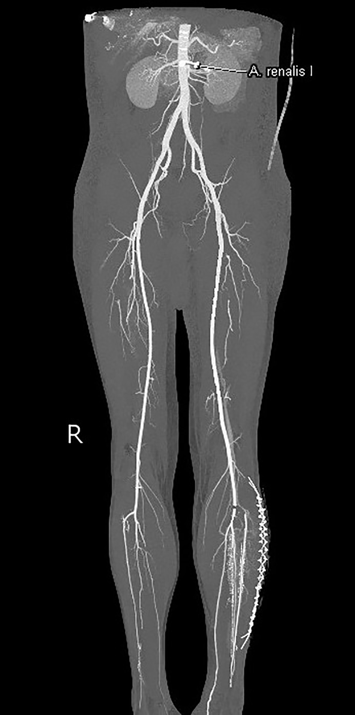
Figure 1. Computed tomography angiography of the abdomen and lower extremities. Successful revascularization with only a minor occlusion at the ipsilateral ostium of the anterior tibial artery.
Figure 2.
Figure 2. Transthoracic ultrasound (apical four-chamber view) shows a dilated right ventricle and right atrium. RV: right ventricle; RA: right atrium; LV: left ventricle; LA: left atrium.
Figure 3.
Figure 3. Computed tomography angiography of the abdomen and thorax. (a) The white arrow shows the deep vein thrombosis on the contralateral femoro-popliteal level. (b, c) Bilateral lung embolization.
Figure 4.
Figure 4. Transesophageal ultrasound with CFM (short-axis view) demonstrates a PFO with a small left-to-right shunt (arrow). IV contrast and Valsalva maneuver also confirmed the presence of a right-left shunt (not recorded). LA: left atrium; RA: right atrium; Ao: proximal ascending aorta.