Journal of Medical Cases, ISSN 1923-4155 print, 1923-4163 online, Open Access
Article copyright, the authors; Journal compilation copyright, J Med Cases and Elmer Press Inc
Journal website http://www.journalmc.org

Case Report

Volume 8, Number 9, September 2017, pages 277-279


Multifocal Ancient Thoracic Schwannomas: A Case Report and Review of Literature

Figures

Figure 1.
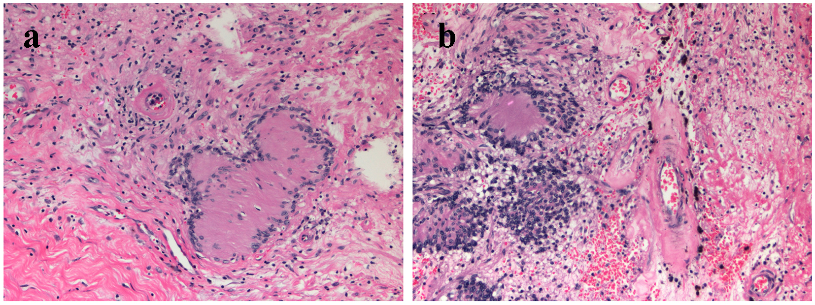
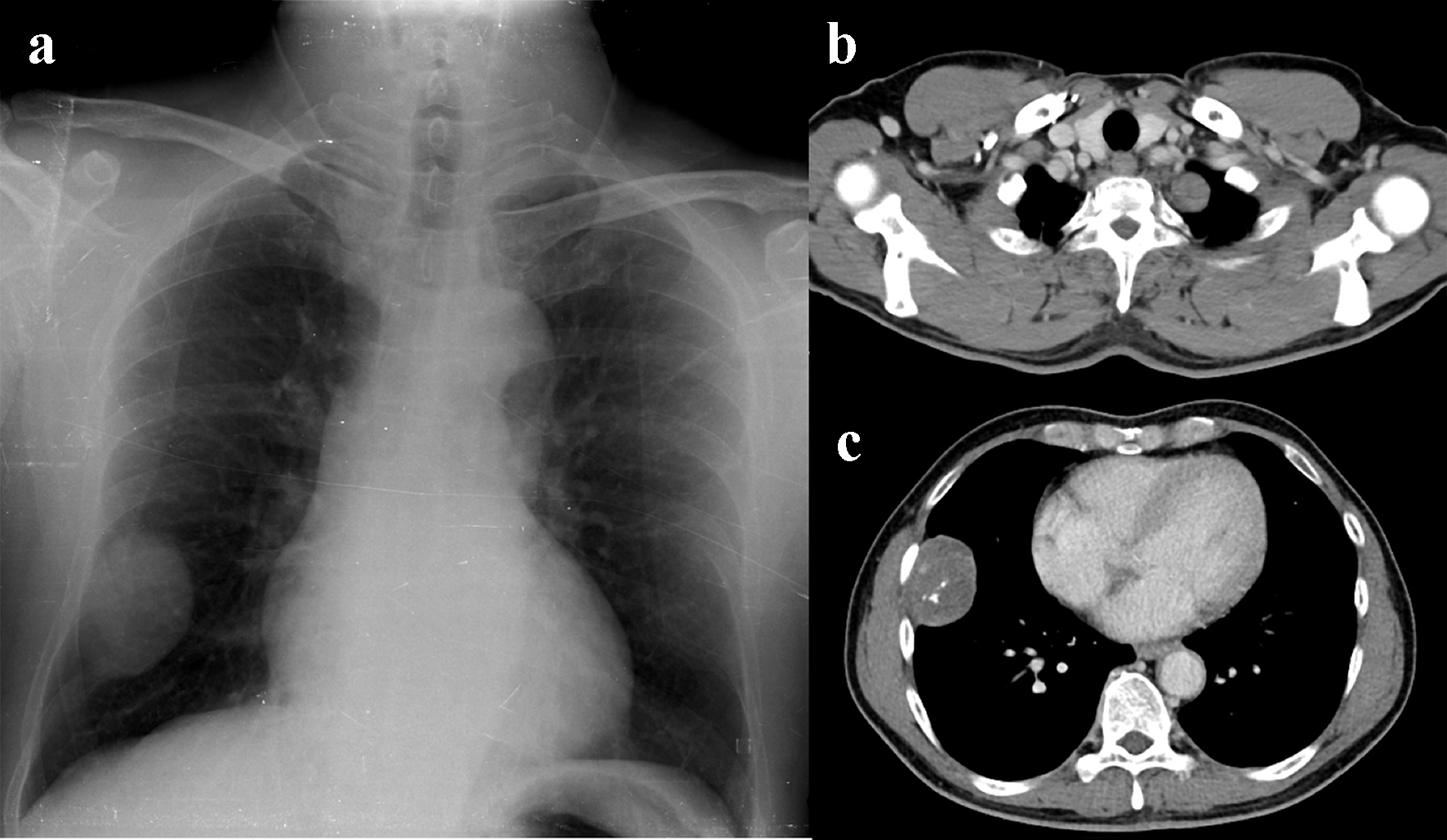
Figure 1. (a) Chest radiography showing a well-circumscribed pleural-based mass containing several small speckled calcifications along the lateral aspect of the right lower hemithorax. Also note the smaller subtle pleural-based lesion at the left apical thorax. (b, c) Axial contrast chest MDCT scan showing two slightly hypodense pleural lesions with mild enhancement at the left apical thorax (b) and right lateral lower hemithorax (c). A few small internal speckled calcifications are noted in the larger one. No adjacent rib destruction or evidence of chest wall invasion was detected.
Figure 2.
Figure 2. (a) Microscopic picture (hematoxylin and eosin staining, × 200) showing Verocay body, formed by alternating rows of palisading nuclei and intervening nuclei-free stroma. (b) Microscopic picture (hematoxylin and eosin staining, × 200) showing degenerative changes including hemorrhage, and hyalinization of blood vessels.