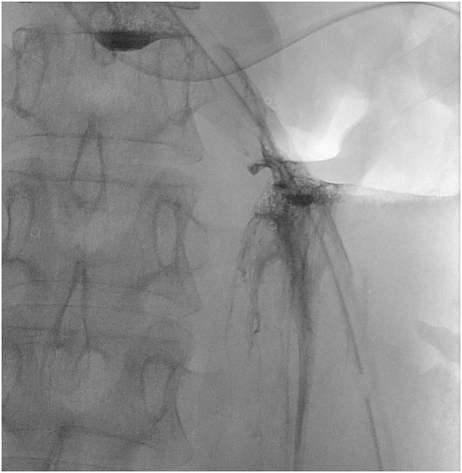
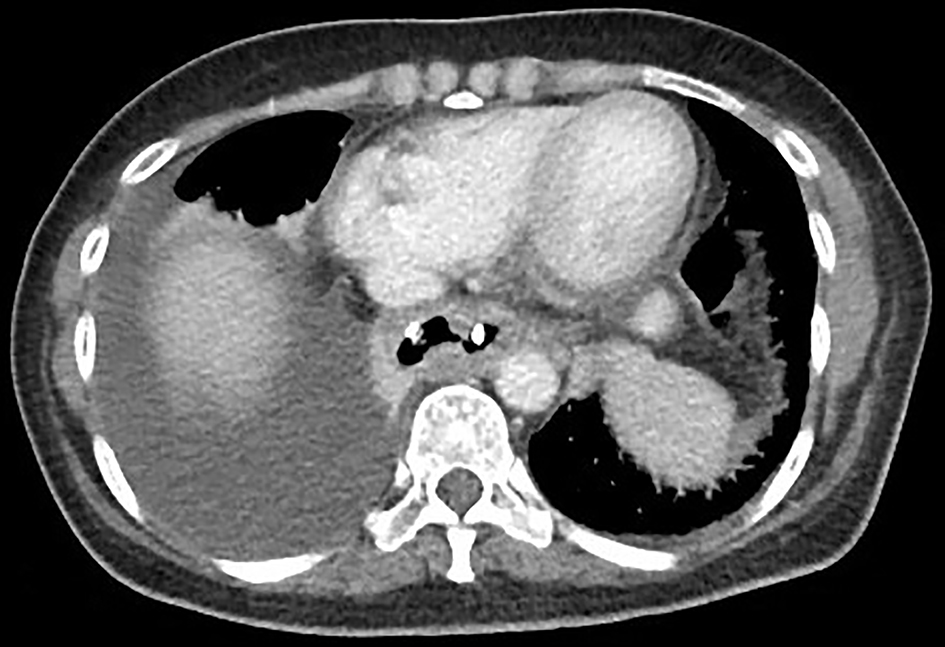
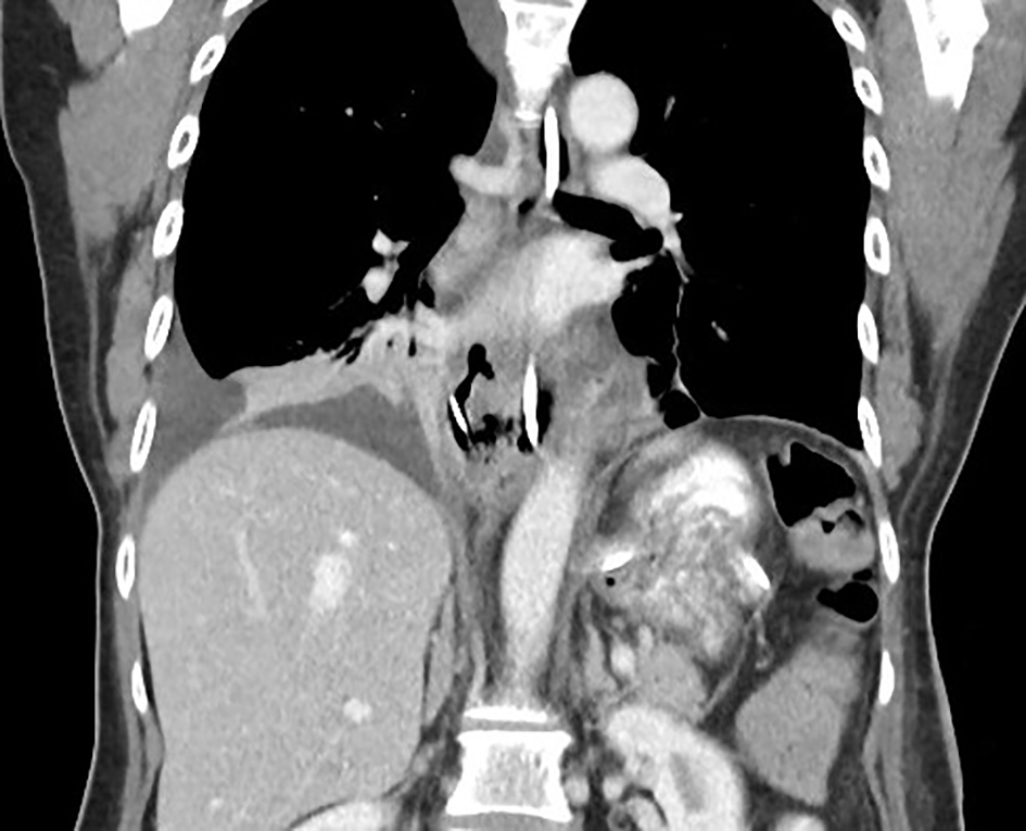
Figure 1. Gastrografin swallow series showing esophageal leak.
| Journal of Medical Cases, ISSN 1923-4155 print, 1923-4163 online, Open Access |
| Article copyright, the authors; Journal compilation copyright, J Med Cases and Elmer Press Inc |
| Journal website http://www.journalmc.org |
Case Report
Volume 8, Number 7, July 2017, pages 215-218
Laparoscopic Enucleation of Schwannoma Masquerading as a Leiomyoma
Figures
Table
| Author | Lesion(s) size | Lesion location | Surgical approach |
|---|---|---|---|
| Shichinohe et al (2014) (case 1) [4] | 50 × 20 mm | Mid esophagus | Right thoracoscopic enucleation |
| Shichinohe et al (2014) (case 2) [4] | 45 × 30 mm | Lower esophagus | Left thoracoscopic enucleation |
| Jeon et al (2014) (case 1) [3] | 95 × 70 × 65 mm + 88 × 50 × 55 mm | Upper esophagus | Right thoracotomy enucleation |
| Jeon et al (2014) (case 2) [3] | 60 × 58 × 40 mm | Upper esophagus | Left cervical approach enucleation |
| Makino et al (2013) [5] | 22 × 34 × 29 mm | Upper esophagus | Right thoracoscopic enucleation |
| Liu et al (2013) [6] | 90 × 90 mm | Mid esophagus | Ivor Lewis partial esophagectomy |
| Kitada et al (2013) [7] | 75 × 57 × 80 mm | Mid esophagus | Left thoracoscopic enucleation |
| Kassis et al (2012) [8] | 113 × 84 × 58 mm | Upper esophagus | three-field esophagogastrectomy + cervical esophagogastric anastomosis |
| Wang et al (2011) [9] | 55 × 40 × 45 mm | Lower esophagus | Left thoracotomy enucleation |
| Dutta et al (2009) [20] | 90 × 60 mm + 60 × 50 mm | Mid esophagus + lower esophagus | Right posterolateral thoracotomy enucleation + trans-thoracic esophagectomy with gastric pull-up and cervical esophago-gastric anastomosis |
| Retrosi et al (2009) [10] | 40 × 60 mm | Upper esophagus | Right cervical approach enucleation |
| Matsuki et al (2009) [11] | 40 × 30 × 35 mm | Upper esophagus | Right thoracotomy enucleation |
| Zhang et al (2008) [12] | 80 × 75 × 50 mm | Upper esophagus | Enucleation - technique unspecified |
| Yoon et al (2008) [13] | 70 × 60 × 40 mm | Upper esophagus | Enucleation - technique unspecified |
| Mizuguchi et al (2008) [14] | 80 × 75 × 60 mm | Upper esophagus | Axillary thoracotomy + VATS enucleation |
| Tokunaga et al (2007) [15] | 74 × 56 × 22 mm | Upper esophagus | Right axillary thoracotomy enucleation |
| Park et al (2006) [16] | 150 × 150 × 45 mm | Upper esophagus | Total thoracic esophagectomy + cervical esophagogastrostomy, pyloroplasty, and feeding jejunostomy |
| Marin et al (2006) (case 1) [17] | Size unspecified | Upper esophagus | Cervical approach enucleation |
| Marin et al (2006) (case 2) [17] | 55 mm | Upper esophagus | Cervical approach enucleation |
| Chen et al (2006) [18] | Size unspecified | Upper esophagus | Cervical approach enucleation |
| Basoglu et al (2006) [19] | 60 × 60 | Upper esophagus | Abdominocervical approach subtotal esophagectomy + esophagogastrostomy |