Journal of Medical Cases, ISSN 1923-4155 print, 1923-4163 online, Open Access
Article copyright, the authors; Journal compilation copyright, J Med Cases and Elmer Press Inc
Journal website http://www.journalmc.org

Case Report

Volume 8, Number 4, April 2017, pages 127-131


Synchronous Infrasellar and Cervical Chondroid Chordoma: A Case Report and Review of the Literature

Figures

Figure 1.
Figure 1. Magnetic resonance imaging (MRI) of solid expansive infrasellar and cervical process with high enhancement post-intravenous injection of paramagnetic contrast. (a) T2 sequence axial view. (b) T1 fat sat and gadolinium sagittal view. (c) T1 fast sat and gadolinium axial view.
Figure 2.
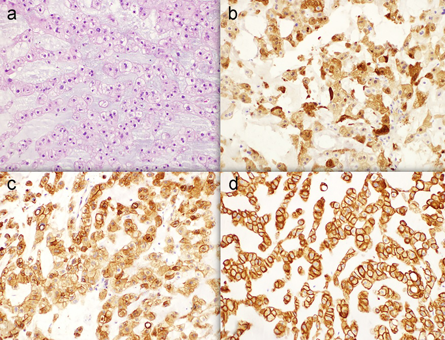
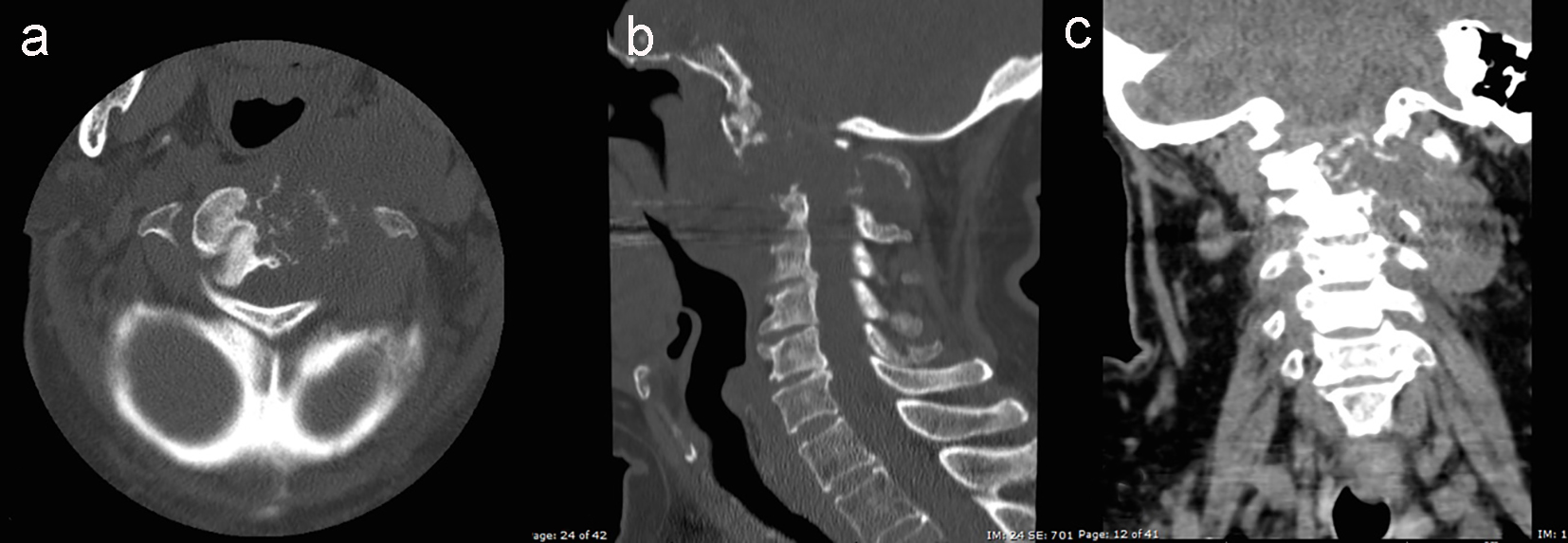
Figure 2. Computed tomography (CT) of expansive and destructive epidural spinal space process, centered in C2 with involvement of C1 and C3 and infiltrative compromise of neighboring left soft tissues. High enhancement after intravenous contrast injection was observed. (a) Bone window axial view. (b) Bone window sagittal view. (c) Soft tissues window and intravenous contrast, coronal view.
Figure 3.
Figure 3. Immunohistochemistry study (× 20). (a) Hematoxylin and eosin stain. (b) Positive S-100 stain. (c) Positive EMA stain. (d) Positive KAE1/3 stain.
Figure 4.
Figure 4. Three-dimentional conformal radiotherapy plan.
Figure 5.
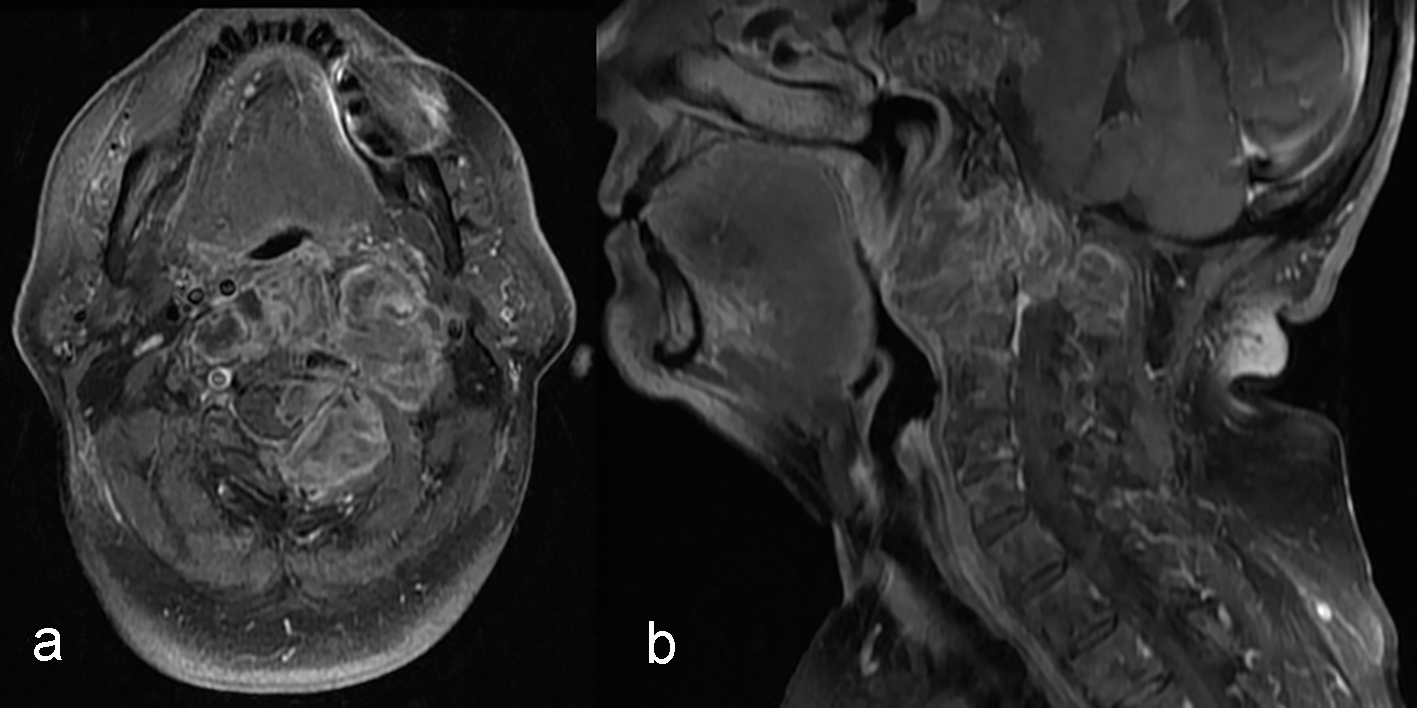
Figure 5. Magnetic resonance imaging (MRI) control imaging after palliative radiotherapy treatment showing growing of the expansive cervical spine process. The mass involved parapharyngeal, retropharyngeal, pharyngeal mucous space and perivertebral spaces, trespassing the middle line and involving the great vessels of the suprahyoid neck. (a) T1 fast sat and gadolinium axial view. (b) T1 fat sat and gadolinium sagittal view.