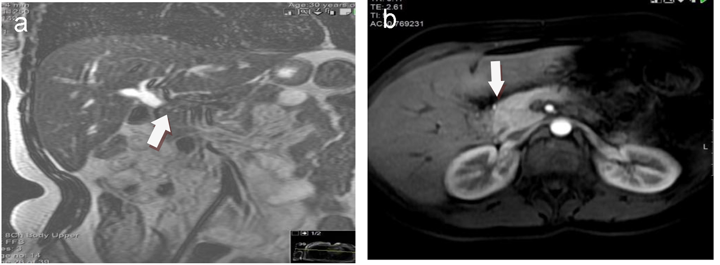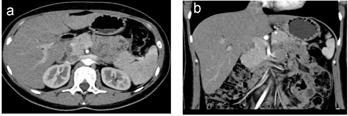
Figure 1. (a) Axial CT and (b) coronal reconstruction images show that there is no any visible mass.
| Journal of Medical Cases, ISSN 1923-4155 print, 1923-4163 online, Open Access |
| Article copyright, the authors; Journal compilation copyright, J Med Cases and Elmer Press Inc |
| Journal website http://www.journalmc.org |
Case Report
Volume 8, Number 4, April 2017, pages 132-136
Granular Cell Tumor of the Common Bile Duct: A Case Report of a Rare Anatomical Entity and Brief Review of the Literature
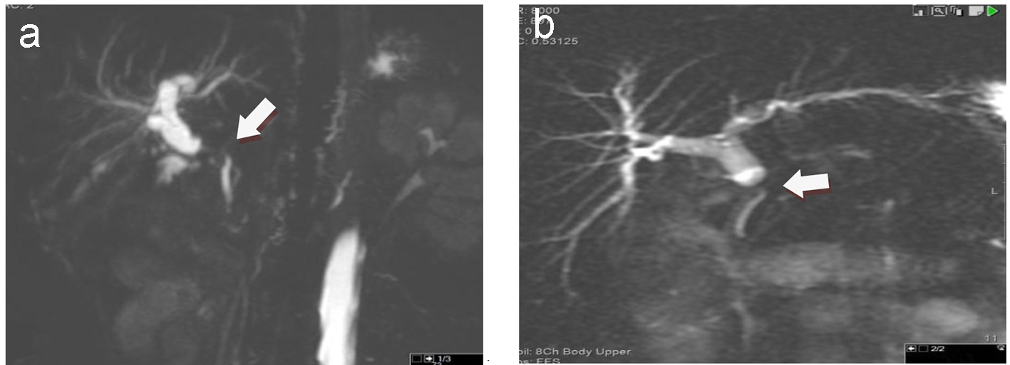
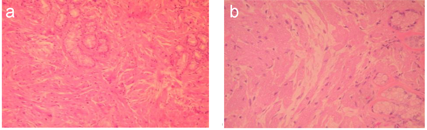
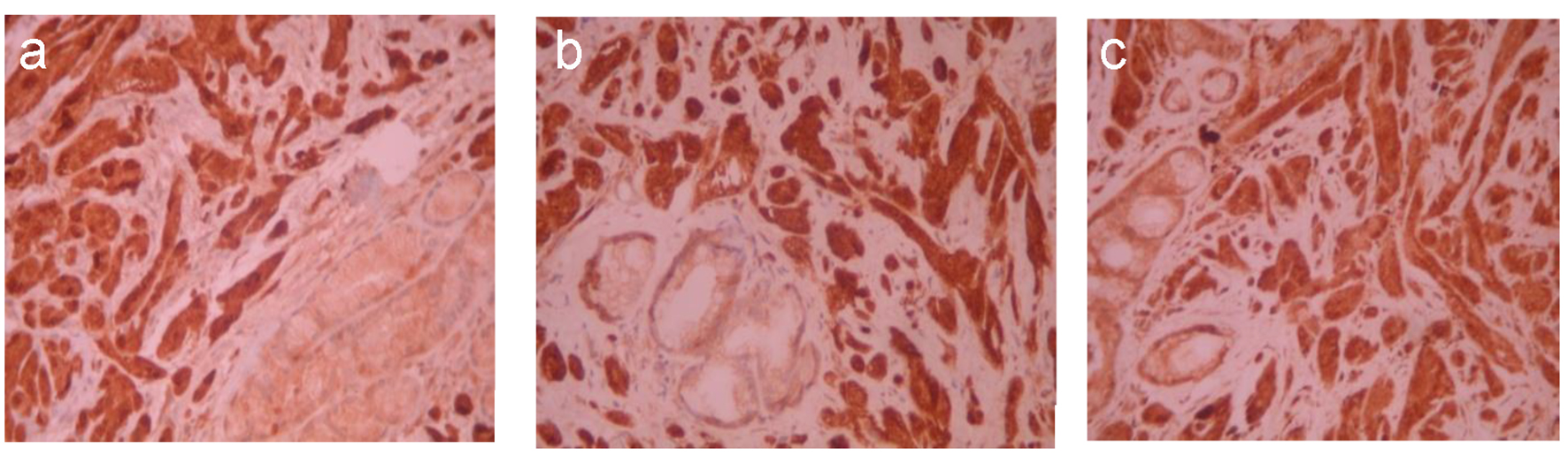
Figures