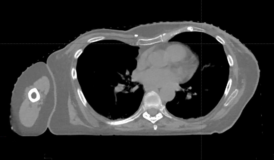
Figure 1. This patient’s chest deformity consistent with PE. Medial axial thoracic CT scan demonstrates the compression of heart and anterior mediastinal structures due to inward sunken sternum and costal-cartilages.
| Journal of Medical Cases, ISSN 1923-4155 print, 1923-4163 online, Open Access |
| Article copyright, the authors; Journal compilation copyright, J Med Cases and Elmer Press Inc |
| Journal website http://www.journalmc.org |
Case Report
Volume 8, Number 3, March 2017, pages 98-101
Advantages of Post-Mastectomy Proton Beam Therapy in a Breast Cancer Patient With Pectus Excavatum
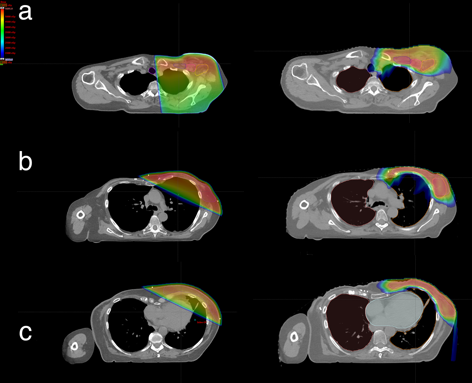
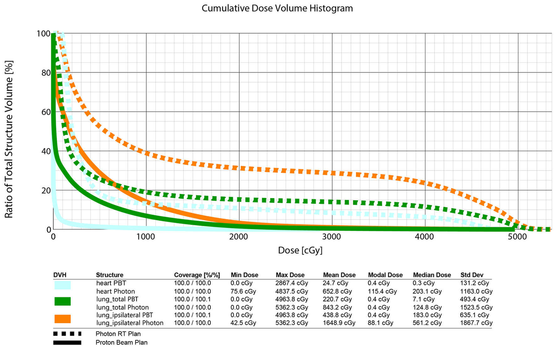
Figures