Journal of Medical Cases, ISSN 1923-4155 print, 1923-4163 online, Open Access
Article copyright, the authors; Journal compilation copyright, J Med Cases and Elmer Press Inc
Journal website http://www.journalmc.org

Case Report

Volume 7, Number 12, December 2016, pages 539-542


Primary T-Cell Lymphoma With Small Intestinal Perforation: A Case Report

Figures

Figure 1.
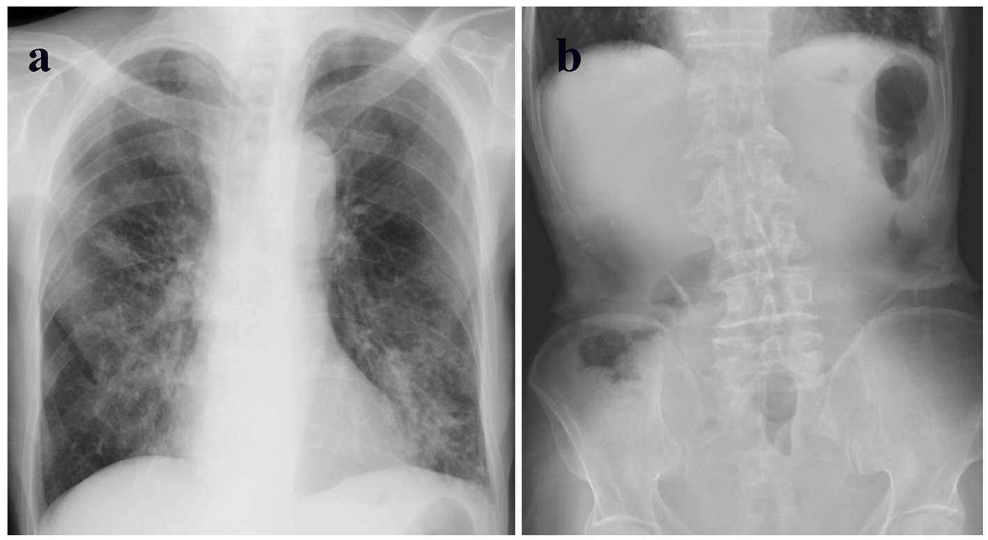
Figure 1. (a) Chest X-ray showing infiltration shadows in bilateral lower lungs. (b) Abdominal X-ray showing the absence of free air.
Figure 2.
Figure 2. (a) Computed tomography showing mild infiltration shadows in the bilateral lower lungs. (b) A small amount of free air on the surface of the left lobe of the liver. (c) Thickened jejunal walls. (d) Enlarged lymph nodes around the thickened jejunum.
Figure 3.
Figure 3. (a) Perforation site located in the upper jejunum (indicated by a circle) with a tumor and several enlarged lymph nodes around the lesion (indicated by a square). (b) Gross specimen of the resected tumors is shown. (c, d) Microscopic specimens stained by hematoxylin-eosin showing massive invasion of large-sized atypical lymphatic cells (c: low-power field; d: high-power field).
Figure 4.
Figure 4. (a, b, and c) Immunohistochemical staining of tumor specimen showing cells positive for CD45 (a) and CD3 (b) and negative for CD20 (c). (d) Immunohistochemical staining for Ki-67 showing 50% of cancer cells as positive.