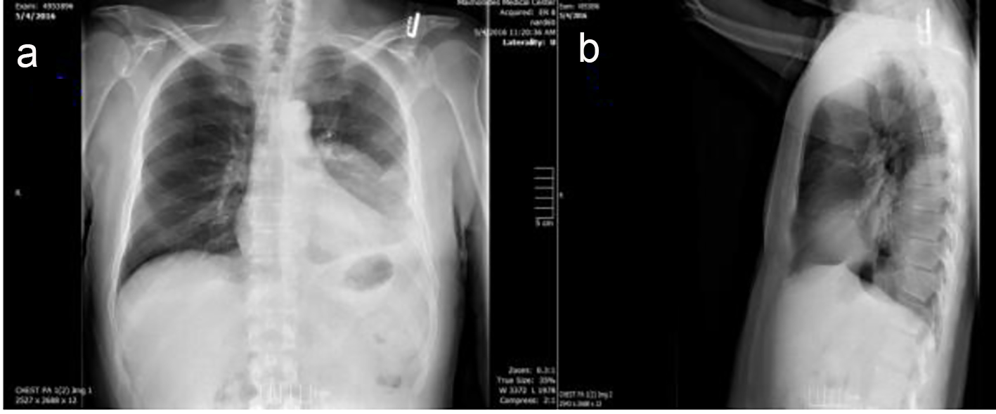
Figure 1. (A) Posterior-anterior and (B) lateral views on initial chest X-ray with findings of left pleural effusion and left lower lobe infiltrate.
| Journal of Medical Cases, ISSN 1923-4155 print, 1923-4163 online, Open Access |
| Article copyright, the authors; Journal compilation copyright, J Med Cases and Elmer Press Inc |
| Journal website http://www.journalmc.org |
Case Report
Volume 7, Number 11, November 2016, pages 512-514
Empyema and Cavitation After Dental Procedure
Figures