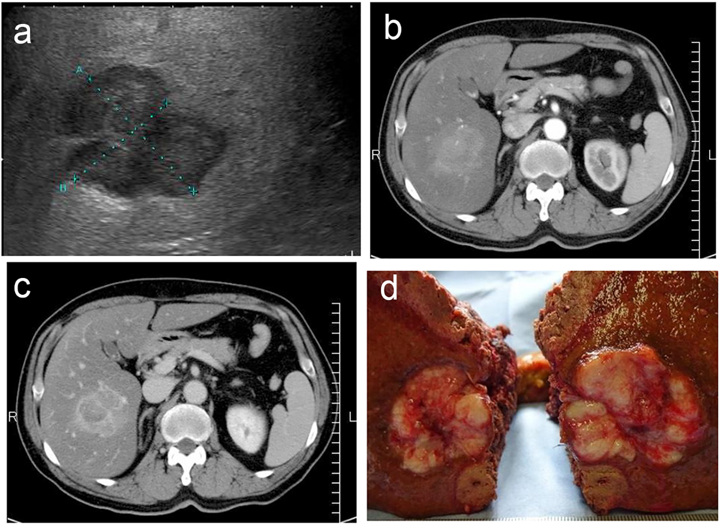
Figure 1. (a) Ultrasonography revealed a lobular mass in S5 of the liver with a marginal low echo, measuring 50 × 40 mm. (b) Contrast-enhanced CT revealed a mass in S5 of the liver, measuring 48 × 45 × 45 mm, with marked enhancement in the early phase. (c) Contrast agents in interiors of the mass were washed out in the late phase with the residues of the contrast agent at the margin of the tumor. (d) Gross specimen revealed a white-yellowish soft tumor with multiple nodules.
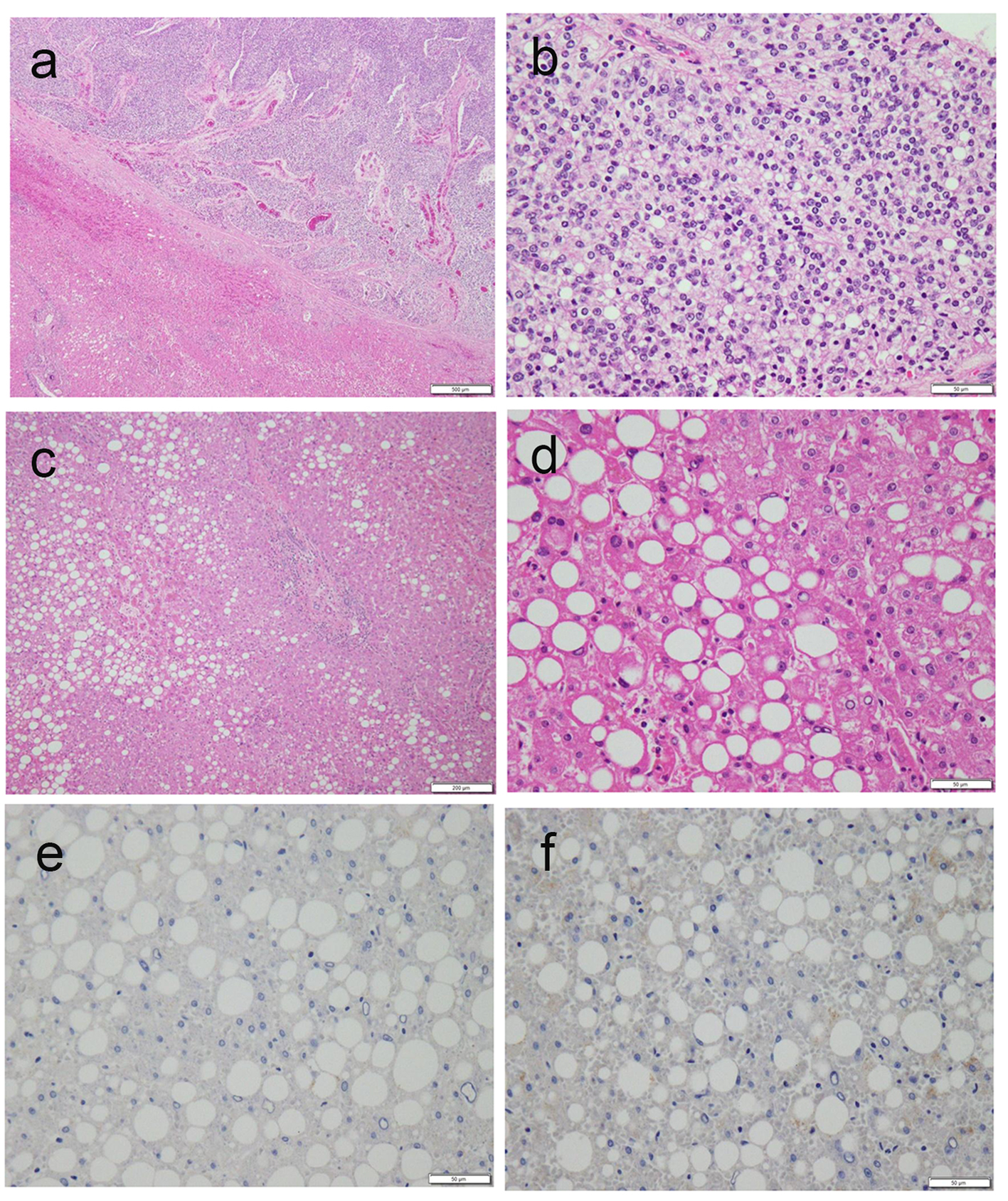
Figure 2. (a, b) Microscopic histological findings of the tumor revealed moderately differentiated HCC (a, low power fields; b, high power fields). (c, d) Macrovesicular lipid depositions and mild steatosis without fibrosis were observed in the background liver, compatible with A2F1 Inuyama classification (c, low power fields; d, high power fields). (e, f) Immunostaining of background liver tissues by HBs-Ag and HBc-Ag revealed negative and false-positive results, respectively.