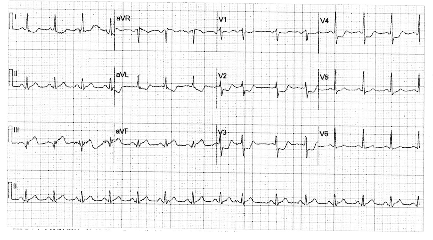
Figure 1. Initial 12-lead electrocardiogram showing ST segment elevation in the inferior leads and reciprocal ST segment depression in the precordial leads.
| Journal of Medical Cases, ISSN 1923-4155 print, 1923-4163 online, Open Access |
| Article copyright, the authors; Journal compilation copyright, J Med Cases and Elmer Press Inc |
| Journal website http://www.journalmc.org |
Case Report
Volume 7, Number 12, December 2016, pages 527-530
Spontaneous Vanishing of Coronary Collaterals During Cardiac Catheterization for Acute Myocardial Infarction
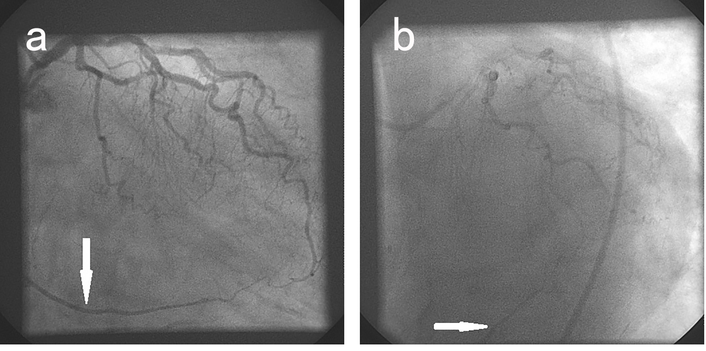
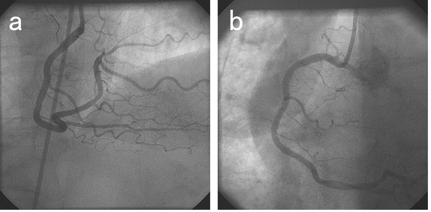
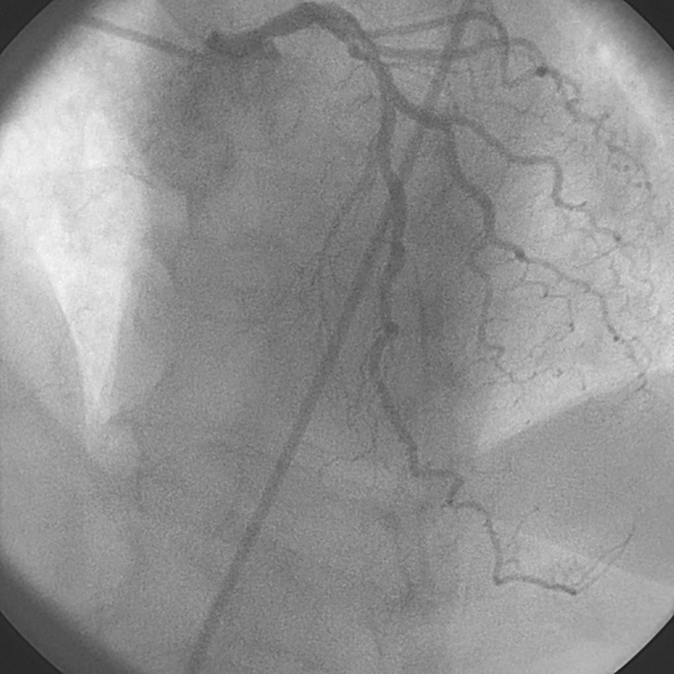
Figures