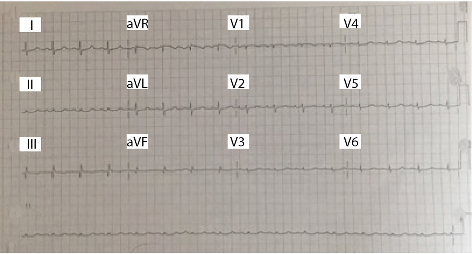
Figure 1. EKG with normal sinus rhythm, normal axis, no ST or ischemic changes, low voltage criteria present.
| Journal of Medical Cases, ISSN 1923-4155 print, 1923-4163 online, Open Access |
| Article copyright, the authors; Journal compilation copyright, J Med Cases and Elmer Press Inc |
| Journal website http://www.journalmc.org |
Case Report
Volume 7, Number 11, November 2016, pages 498-501
A Rare but Lethal Malignant Cardiac Mass
Figures