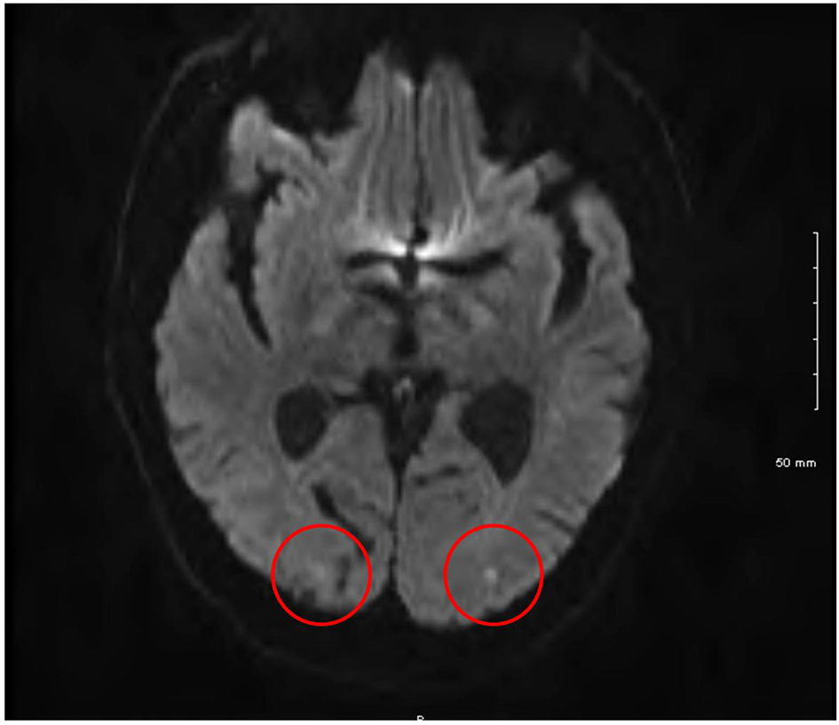
Figure 1. Cranial magnetic resonance imaging showing bilateral occipital lobe infarcts.
| Journal of Medical Cases, ISSN 1923-4155 print, 1923-4163 online, Open Access |
| Article copyright, the authors; Journal compilation copyright, J Med Cases and Elmer Press Inc |
| Journal website http://www.journalmc.org |
Case Report
Volume 7, Number 9, September 2016, pages 379-383
A Case of Transient Loss of Vision Following Coronary Angiography: Etiology, Investigation and Management
Figures
Table
| Cerebrovascular disease secondary to embolism (thrombus or atheroma), in situ thrombosis or intracerebral hemorrhage. |
| Catheter-related vasospasm of cerebral vessels. |
| Intimal tears causing dissection of the aortic arch and its branches. |
| Contrast-induced cortical blindness. |
| Hypotension, which may be contrast-induced. |
| Hypoventilation. |
| Migraine. |
| In addition, cortical blindness has been observed in patients following head trauma and in those with uremia, meningitis and hysteria. |