Journal of Medical Cases, ISSN 1923-4155 print, 1923-4163 online, Open Access
Article copyright, the authors; Journal compilation copyright, J Med Cases and Elmer Press Inc
Journal website http://www.journalmc.org

Case Report

Volume 7, Number 8, August 2016, pages 354-357


Epstein-Barr Virus-Induced Polymorphic Nodal Lymphoproliferative Disorder Presenting as Recurrent Fever and Scrotal Swelling: A Case Report and Review of Literature

Figures

Figure 1.
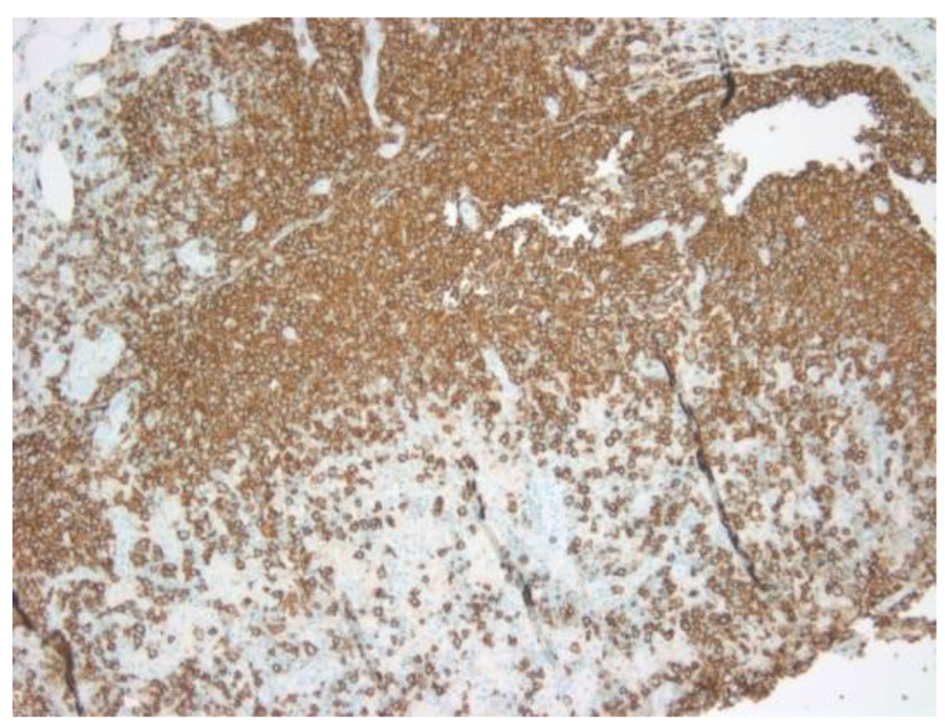
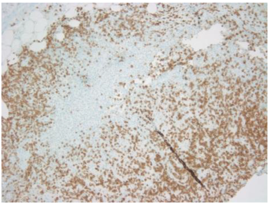
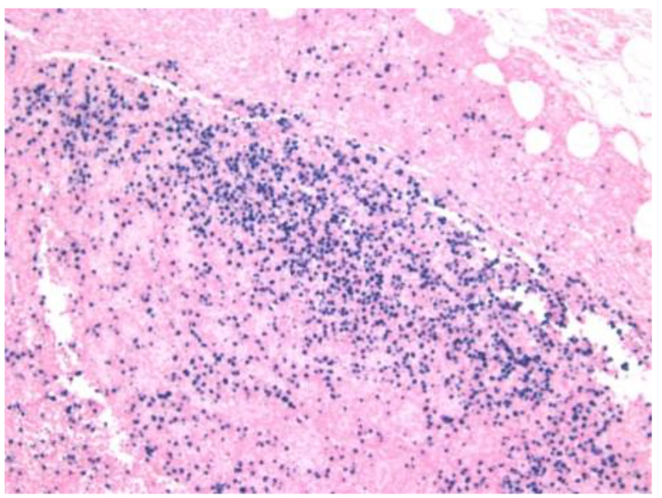
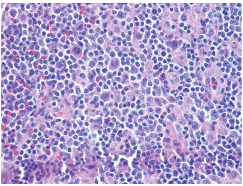
Figure 1. H&E sections reveal lymph node with an altered architecture with vague nodularity, separated by fibrotic bands, and lymphoid infiltration into the perinodal adipose tissue. Nodules are composed of mixed inflammatory infiltrates consisting of polymorphous infiltrate comprising small lymphocytes, histiocytic aggregates, large atypical cells, immunoblasts, mummified cells, neutrophils/eosinophils and plasma cells, rare atypical RS type cells and increased vascularity comprised of high endothelial vessels.
Figure 2.
Figure 2. CD20 showing atypical large B cells.
Figure 3.
Figure 3. CD3 showing scattered positivity in atypical large T cells.
Figure 4.
Figure 4. EBV in situ hybridization positive in B and T cells.